- Physiologically realistic morphological, fluidic and 3D cellular conditions
- Universal platform with architecture specific of desired organ
- Significant reduction in cost and time
- Robust and easy to use protocols
- Compatible with standard analytical instruments for both on chip and off chip assays including omic methodologies for systems biology and bioinformatic analysis
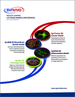
SynTox is the only commercially available 3D toxicology model with real-time optical monitoring and multi-compartment, multi-cellular architecture and low reagent requirements. Other benefits of this platform are:

SynTox 3D Toxicology Model recreates the in vivo micro-environment by emulating a histological slice operating in an in vitro Environment.
Current in vitro platforms are poor predictors of in vivo safety, efficacy and pharmacokinetics of therapeutics owing to significant difference in the test conditions compared to physiological conditions observed in vivo.
)window.location='https://i2.wp.com/www.synvivobio.com/wp-content/uploads/2016/07/invitro-invivo.jpg')
Current in vitro models routinely utilize 2D monolayers or 3D aggregates of cells under static conditions for studying drug toxicity. These models fail to reproduce in vivo physiological features such as morphological size, physiological blood flow and cellular (biological) make-up of the specific organs being investigated. Other microfluidic models employ a membrane-based top-bottom two-compartment architecture, inherently limiting key desired features such as real-time visualization and the ability to simultaneous analyze multi-cellular cultures.
SynTox 3D Model Kit Components
All the basic components required to run the SynTox assay can be purchased in a kit format. All accessories including tubing, clamps, needles and syringes are included. Starter kits will also include the pneumatic priming device (required for running SynTox assays).
| Qty | Description | Cat# |
|---|---|---|
| 10 | SynTox Chips | 102016 |
| 100ft | Tygon Tubing (0.02″ ID x 0.06″ OD) | 201005 |
| 25 | Slide Clamps | 202003 |
| 50 | Blunt Tip Needles (24ga x 0.5″ long) | 204002 |
| 50 | 1mL Syringes with Luer-Lock Tips | 203004 |
| 1 | SynVivo Pneumatic Primer Device (in starter kits only) | 205001 |
SynTox Liver model was used to demonstrate toxicity of classical pain killer acetaminophen using different modes of interaction.
Hepatocytes were cultured in SynTox model and analyzed for functionality using standard commercially available assays. Hepatocytes formed bile-canaliculi, produced urea in increasing concentration in time dependent manner with upregulated enzyme activity under physiological fluid flow conditions.
)window.location='https://i1.wp.com/www.synvivobio.com/wp-content/uploads/2016/07/syntox-liver1.jpg')
Hepatocytes form bile-canaliculi (Figure A), secrete urea with increased production in time dependent manner (Figure B) and can be monitored in real-time for enzyme activity (Figure C). Effect of flow on hepatocytes is clearly observed with increased enzyme activity.
SynTox Toxicology Model was used for understanding of drug toxicity responses using systems biology based analysis.
Liver cells and heart cells were co-cultured with their respective endothelial cells in the SynTox microfluidic chip and treated with Doxorubicin. The cells were harvested and subjected to genomic analysis resulting in upregulated and downregulated genes. The identified genes were used to develop systems biology based cellular pathway model for identification of targets and mechanism.
)window.location='https://i2.wp.com/www.synvivobio.com/wp-content/uploads/2016/07/drug-toxicity-response-genomics.png')
Genomic responses highlighting upregulated and down regulated cells in Endothelial Cells (Figure A), Hepatocytes (Figure B) and Cardiac Cells (Figure C)
Mono cultured hepatocytes or co-cultured with endothelial cells were treated with acetaminophen and their drug responses were investigated for different modes of interactions using a combination of Live/Dead and Reactive Oxygen Species (ROS) assays. Differences between vascular and direct treatment of hepatocytes was observed following drug interaction.
)window.location='https://i2.wp.com/www.synvivobio.com/wp-content/uploads/2016/07/syntox-liver2.jpg')
Viable co-cultures of endothelial cells and hepatocytes were observed in SynTox model(Figure A). C. Co-Culture of endothelial cells and liver cells were treated with a dose of acetaminophen followed by wash and continuous perfusion of fresh media. As seen from the images, the liver cells in the periphery (Figure D) are prone to death compared to the cells away from the vasculature as they take up most of the drugs. In contrast, none of the endothelial cells are showing any signs of distress indicating that they are not affected by the drug. Static treatment with drug results in maximum cell death (Figure C). Comparison of different modes of drug treatment for the same dosage and duration (Figure D). In contrast to static systems that over predict the toxicity responses, SynVivo reproduces the in vivo therapeutic responses.
)window.location='https://i0.wp.com/www.synvivobio.com/wp-content/uploads/2016/07/real-time-monitoring.png')