����3D����������Է����ǣ�3D THICKNESS MAPPING���ؽڱ���������ͼ�����ǣ��ؽ����ǵ�ѹ����������ؽ����ǵ�ѹ��ʵ��ϵͳ
�ͺ�:3D THICKNESS MAPPING
��ϵ��:������
��ϵ�绰:18618101725
Ʒ��:BMM
����3D����������Է����ǣ�3D THICKNESS MAPPING���ؽڱ���������ͼ������
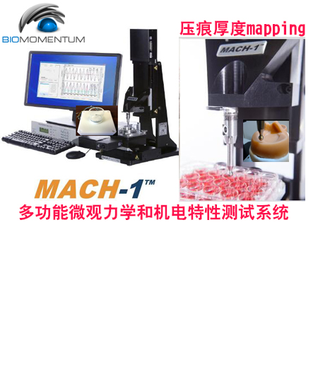
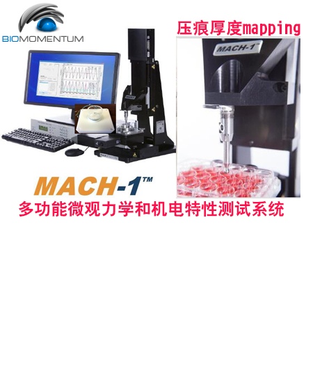
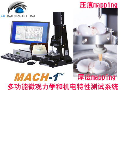
ʹ�ò����������·�������̽��3D���ӳ�䣬�����ڽ�Ӳ�Ļ��ģ����磬��ͷ�ϵ����Dz㣩�ϻ��Ʋ��ϵı��������ͺ�ȡ� ����ͨ���� XYɨ�衱�͡����ҽӴ���������ɵġ� �����ҽӴ��������Ժ㶨�������ƶ���ͷ��ֱ���������ϲ����ϣ��������ǣ����棬���ڼ����ɵ���Ӳ�IJ��ϣ����������ʱֹͣ�� ����Ĵ�ֱλ�ã��غɿ�ʼ���ӵ�λ�ã������Ĵ�ֱλ�ã�λ��/�������еĸ��յ㣩֮��IJ��ṩ�˴�ֱ��ȡ� ����ʹ����ǰʹ��MachOne Motion�����еġ�����ѹ�ۡ����ܻ�õı��淽���������ȷֲ���
- ֮3D����ѹ��ӳ��ͺ�Ȳ��Է�����3D NORMAL INDENTATION MAPPING�� ��3D���ӳ����Է�����3D THICKNESS MAPPING��
ѹ�������ص�
1���ɲ���������ѹ��mapping�ͺ��mapping
2�����ڶ�װ��и�ͨ��ѹ�۲��Ժͺ�Ȳ���
3������������Һ��ֱ��ѹ�۲��Ժͺ�Ȳ���
4����Ҫ��ֱѹ��
5��ѹ�ۺͺ���Զ�����������
6����Ʒ���죬������������ƪ��
������ף�
Sensitivity of indentation testing to step-off edges and interface integrity in cartilage repair
��������̨�ױ�Ե�ͽ���������ѹ�۲���������
Indentation probing of human articular cartilage: effect on chondrocyte viability
����ؽ����ǵ�ѹ��̽�⣺������ϸ��������Ӱ��
Assessment of Human Articular Cartilage Issued from Asymptomatic & TKR DonorsHadjab I, Sim S, Quenneville E, Garon M and Buschmann MDBiomedical Engineering Society 2015 in Tampa, Floride
Introduction: Cartilage degeneration is a progressive process and currently, only end-stage surgical treatments such as total knee replacement (TKR) lead to an improved condition. To prevent or delay this surgery, several less invasive alternatives could be considered, such as pharmacological treatments, scaffolds and partial replacements1. However, current diagnostic techniques are limited...Read More
Micro-CT visualization and indentation properties of whole meniscus following mercury exposureKolaczek S, Changoor A, Hurtig M, Gordon K and Getgood AAnnual Meeting of the ORS, New Orleans, Louisiana, USA, March 15-18, 2014.
A major risk factor for knee osteoarthritis (OA) is meniscal injury or excision. Surgeons now advocate for meniscal repair or allografts rather than excision with this knowledge in hand. Clinical assessment of the meniscal load sharing is limited to meniscus position by MRI, so the efficacy of replacement and repair must be done in cadaveric knees. In the past, pressure sensitive film and mechanical...Read More
Mapping Articular Cartilage Biomechanical Properties of Normal and Osteoarthritis Mice Using IndentationLavoie JF, Sim S, Quenneville E, Garon M, Moreau A, Buschmann MD and Aubin CE2015 International Cartilage Repair Society, May 7-11 2015, Chicago, Illinois, USA. Podium presentation (ID: 23.2.9)
Purpose: Due to their size (~1mm), mouse models pose significant challenges to map biomechanical properties over their articular surfaces. The purpose of this study was to determine if an automated indentation technique could be used to map the biomechanical properties of the articular surfaces in murine knees and to identify early alterations of the articular cartilage of a mouse strain (STR/ort)...Read More
Mapping Articular Cartilage Biomechanical Properties of Normal and Osteoarthritic Mice Using Indentation (OARSI Poster)Lavoie JF, Sim S, Quenneville E, Garon M, Moreau A, Buschmann MD and Aubin CEOsteoarthritis Research Society International (OARSI), 2015 April 30, Seattle, WA, United States
Purpose: Due to their size (~1mm), mouse models pose significant challenges to map biomechanical properties over their articular surfaces. The purpose of this study was to determine if an automated indentation technique could be used to map the biomechanical properties of the articular surfaces in murine knees and to identify early alterations of the articular cartilage of a mouse strain...Read More
Mapping Articular Cartilage Biomechanical Properties of Normal and Osteoarthritic Mice Using IndentationLavoie JF, Sim S, Quenneville E, Garon M, Moreau A, Buschmann MD and Aubin CEOsteoarthritis and Cartilage 23 Suppl. 2, p. A254 (2015)
Purpose: Due to their size (~1mm), mouse models pose significant challenges to map biomechanical properties over their articular surfaces. The purpose of this study was to determine if an automated indentation technique could be used to map the biomechanical properties of the articular surfaces in murine knees and to identify early alterations of the articular cartilage of a mouse strain (STR/ort)...Read More
Contrast-Enhanced Computed Tomography Reflects Stiffness of Intact Articular CartilageNickmanesh R, Stewart R, Snyder B, Grinstaff M and Wilson DISMRM Workshop on Imaging Based Measures of Osteoarthritis. Sept 11-14, 2015. Pacific Grove, CA, USA. Presentation on Sept 13 at 14:39.
Purpose: Articular cartilage distributes load in joints and provides a low-friction surface for joint movement. Glycosaminoglycan (GAG) in cartilage plays a critical role in its compressive stiffness. Loss of GAG is an early sign of osteoarthritis that leads to lower compressive stiffness and altered viscoelastic behavior. MRI and CT-based imaging techniques have been developed to quantify GAG...Read More
Growth Factor Stimulation Improves the Structure and Properties of Scaffold-Free Engineered Auricular Cartilage ConstructsRosa RG, Joazeiro PP, Bianco J, Kunz M, Weber JF, et alPLoS ONE 9(8): e105170. doi:10.1371/journal.pone.0105170. (2014)
The reconstruction of the external ear to correct congenital deformities or repair following trauma remains a significant challenge in reconstructive surgery. Previously, we have developed a novel approach to create caffold-free, tissue engineering elastic cartilage constructs directly from a small population of donor cells. Although the developed constructs appeared to adopt the structural appearance...Read More
Novel Technique to Map the Biomechanical Properties of Entire Mice Articular Surfaces Using IndentationSim S, Lavoie JF, Moreau A, Quenneville E, Garon M, and Buschmann MDOsteoarthritis and Cartilage, 22(1), 541. World Congress on Osteoarthritis, April 2014, Paris, France.
Purpose: An important measure of articular cartilage function in health and disease is its biomechanical properties. While much research has mouse models of osteoarthritis, the assessment of biomechanical properties in these small joints is quite challenging. We have previously developed novel and easily implemented indentation technique on sheep stifle joints and rat knee joints. The purpose...Read More
Novel Technique to Map the Biomechanical Properties of Entire Articular Surfaces Using Indentation to Identify Degenerated (Osteoarthritis-like) CartilageSim S, Chevrier A, Garon M, Quenneville E and Buschmann MD7th World Congress on Biomechanics, 2014, Boston, MA, USA. Poster 2229
Introduction: A currently unsatisfied need in Arthritis and cartilage research is to assess the function of the entire articular cartilage surface both quantitatively and non-destructively. The objective of this study was to investigate the ability of a novel automatic technique to characterize mechanical properties of entire articular surfaces in indentation in order to rapidly discriminate...Read More
Indentation Method to Map Mechanical Properties of Articular Surface to Identify Degenerated RegionsSim S, Chevrier A, Garon M, Quenneville E and Buschmann MD2014 Annual Meeting of the Biomedical Engineering Society, 2014, San Antonio, TX, USA. Podium presentation (ID: OP-Sat-3-4)
Introduction: The identification and quantitative grading of early degenerated regions over an entire articular surface remains a challenging quest. The objective of this study was to investigate the ability of a novel technique to automatically characterize mechanical properties of entire human articular surfaces in indentation in order to rapidly and non-destructively discriminate between...Read More
Relevance of the Spatial Distribution of Mechanical Properties of Articular Cartilage in Animal StudiesSim S, Hadjab I, Garon M, Quenneville E, and Buschmann MDOrthopeadic Research Society Annual Meeting in Las Vegas, 2015, Poster 0359
Introduction: In cartilage regeneration and repair, mechanical testing of articular cartilage characterizes functional restoration of the repair site [1] and can detect early degeneration of articular cartilage [2]. However, the experimental design often incorporates the use of cartilage adjacent to the treated site or at contralateral sites as normal references to evaluate the effect of treatment...Read More
Evaluation of Entire Ovine Cartilage Repair Articular Surfaces: Mechanical and Electromechanical AssessmentSim S, Hadjab I, Garon M, Quenneville E, Hurtig MB, Buschmann MD and Hoemann CDTransactions of International Cartilage Repair Society (ICRS), Chicago, 2015, 7-11 May 2015, e-Poster: P87
Purpose: To demonstrate the ability of non-destructive electromechanical device and automated indentation technique in assessing the quality of cartilage in a sheep model of cartilage repair.
Methods: Ex vivo electromechanical and mechanical mappings of articular surfaces (~40 positions/map) were performed over distal condyles from 5 treated sheep (8 �C 9 y-o,...Read More
Wound Healing Revealed by a Novel Automated Indentation TechniqueSim S, Garon M, Quenneville E and Buschmann MDCanadian Connective Tissue Conference 2015 in Quebec, Canada
Introduction: Mechanical characterization of wound healing in skin samples mostly relies on uniaxial tensile rupture tests, which provide local information along the wound and are disruptive for samples (Chao et al., 2011). In this study, we wanted to test the ability of a novel automated indentation technique to non-destructively characterize mechanical properties of the entire wound and its...Read More
Development of a Sequence of Mechanical Tests for Articular Cartilage at a Single LocationSim S, Chartrand A, Lavallee AP, Tessier J, Garon M, Quenneville E and Buschmann MDOrthopeadic Research Society Annual Meeting in Orlando, 2016
In a recent study, our group has highlighted the importance of considering the natural topographic variability of the mechanical properties over the articular surface, particularly in the context of cartilage repair, where it can screen the effect of a treatment [1]. Moreover, the availability of test sample is limited in those repair studies since the regions of interest are often limited in size....Read More
Cartilage Stiffness and Thickness Distributions Revealed by an Automated Indentation Technique in the Temporomandibular JointSim S, Matuska A, Garon M, Quenneville E, McFetridge P and Buschmann MDTMJ Bioengineering Conference - V, September 12-13, 2016, Barcelona, Spain
The purpose of this study was to evaluate the capability of an automated indentation technique to reveal the topographical variation of mechanical properties over the entire articular surface of the temporomandibular joint (TMJ), especially the thickness and instantaneous modulus (IM). Mechanical properties of visually normal temporal bones and condyles of a porcine TMJ were mapped ex vivo using a...Read More
Correlation of Non-destructive Electromechanical Probe () Assessment with Histological Ss, Biochemical Composition and Mechanical Properties in Human Knee JointsSim S, Chevrier A, Quenneville E, Garon M and Buschmann MDTransactions of the 60th Annual Meeting of the Orthopaedic Research Society, New Orleans, LA, USA, Poster 0439, 2014
Introduction: Histological scoring, biochemical analyses and biomechanical testing (unconfined compression) are often seen as gold standard characterizations for articular cartilage but can present major drawbacks in the context of animal and human studies where characterization of complete intact articular cartilage surfaces is required. In particular, histology, biochemical and mechanical...Read More
Novel Technique to Map the Biomechanical Properties of Entire Articular Surfaces Using Indentation to Identify Osteoarthritis-like RegionsSim S, Chevrier A, Garon M, Quenneville E and Buschmann MDTransactions of the 60th Annual Meeting of the Orthopaedic Research Society, 2014, New Orleans, LA, USA, 2015, Poster #2015
Introduction: It is challenging to identify and grade degenerated regions of the entire articular surface both quantitatively and non-destructively. Therefore, the objective of this study was to investigate the ability of a novel technique to automatically characterize mechanical properties of entire articular surfaces in indentation to rapidly discriminate between damaged articular cartilage...Read More
Evaluation of a novel technique to map the mechanical properties of an entire articular surface in indentationSim S, Quenneville E, Garon M, Hoemann CD, Hurtig M and Buschmann MDInternational Cartilage Repair Society (ICRS), Turkey, 2013, Podium presentation (11.2.9)
Purpose: Mechanical testing of articular cartilage is recommended by the FDA for products intended for the repair or replacement of knee cartilage. One experimental configuration that has many practical advantages is indentation. However, one limitation is the need to perpendicularly position the articular surface to the indenter. The objective of this study was to investigate the ability of...Read More
Ermittlung mechanischer Kennwerte mittels IndentationSeidenstucker MBioNanoMat. 2015; 16 (2-3): 152�C156 DOI 10.1515/bnm-2015-9014
(In Germany only) In der Materialpr��fung sind Indentationsverfahren bereits seit Jahren g?ngige Praxis. Jedoch war es bisher nicht so ohne weiteres m?glich Gewebeproben, insbesondere Weichgewebe wie Knorpel zu untersuchen. Mit dem Mikroindenter MachOneTM von BMM k?nnen sowohl flexible Biomaterialien wie Kontaktlinsen oder Wundauflagen aus elektrogesponnenen Gelatinefliesen genauso wie biologische...Read More
Guidelines for an optimized indentation protocol for measurement of cartilage stiffness - The effects of spatial variation and indentation parametersMoshtagh PR, Pouran B, Korthagen NM, Zadpoor AA and Weinans HJournal of Biomechanics 49(14) �� September 2016 DOI: 10.1016/j.jbiomech.2016.09.020
Mechanical properties of articular cartilage that are vital to its function are often determined by indentation tests, which can be performed at different scales. Cartilage tissue exhibits various types of structural, geometrical, and spatial variations that pose strict demands on indentation protocols. This study aims to define a reproducible micro-indentation protocol for measuring the effective...Read More
ASTM F2451-05 - Standard Guide for in vivo Assessment of Implantable Devices Intended to Repair or Regenerate Articular CartilageASTM International, West Conshohocken, PA, 2010, www.astm.org
Significance and Use
This guide is aimed at providing a range of in vivo models to aid in preclinical research and development of tissue engineered medical products intended for the clinical repair or regeneration of articular cartilage.
This guide includes a description of the animal models, surgical considerations, and tissue processing as well as the qualitative and quantitative...Read More
ASTM F2791 - Standard Guide for Assessment of Surface Texture of Non-Porous Biomaterials in Two DimensionsASTM International, West Conshohocken, PA, 2015, www.astm.org
Significance and Use
4.1 The term ��surface texture�� is used to describe the local deviations of a surface from an ideal shape. Surface texture usually consists of long wavelength repetitive features that occur as results of chatter, vibration, or heat treatments during the manufacture of implants. Short wavelength features superimposed on the long wavelength features of the surface,...Read More
MachOne �C Automated Indentation Mapping (MA056-SOP01-D v2)Sim S and Quenneville EBMM Inc. Laval (QC), Canada, Effective Date: April 7th, 2015
Purpose: This procedure describes a standard method to realize an automated indentation mapping over the surface of a sample using the MachOne mechanical tester (model v500css or v500csst, with software add-on for Automated Indentation Mapping).
Scope: This procedure can be used for the ex vivo automated indentation mapping over the surface of a sample. Sample��s...Read More
MachOne Analysis - Extraction of Elastic Model Parameters Following an Automated Indentation Mapping (SW186-SOP01-D v1)Sim S and Quenneville EBMM Inc. Laval (QC), Canada, Effective Date: April 17th, 2015
Purpose
This procedure describes a method to extract elastic model parameters (Instantaneous modulus, which is referred as the Young��s modulus in MachOne analysis, and shear modulus) from MachOne files generated during an automated indentation mapping of a surface (as per MA056-SOP01-D). It also describes the creation of a corresponding ��.map�� characterization file.
Scope...Read More
Indentation and Thickness Mapping of Articular Surfaces through Manual Positioning (Protocol Template AAAX1 v1)BMM Inc. Laval (QC), Canada, Effective Date: December 6th, 2016.
Purpose
This editable document (MS Word format) is a blank protocol template created to facilitate the creation of your own study protocol involving the indentation and thickness mapping of articular surfaces through manual positioning. It provides suggestions on how to:
-
properly position and secure in place an articular surface into a testing chamber,
-
create...Read More
Combined Mechanical Characterizations Increases Sensitivity in the Assessment of Human Cartilage DegenerationSim S, Hadjab I, Chevrolat L-A, Masse M, Tong AL, Lavigne P, Garon M, Quenneville E and Buschmann MDAccepted for a podium presentation at ORS 2017
Introduction: We published a recent study showing superior sensitivity of electromechanical and indentation (instantaneous response) assessments versus well-established techniques, including histological Mankin s, to characterize cartilage degeneration. This study aims to determine whether the combination of instantaneous, relaxation and equilibrium mechanical properties and friction...Read More
Electromechanical probe and automated indentation maps are sensitive techniques in assessing early degenerated human articular cartilageSim S, Chevrier A, Garon M, Quenneville E, Lavigne P, Yaroshinsky A, Hoemann CD and Buschmann MDJ Orthop Res, 35(4) 858-867. (2017) Epub 2016 Jun 22
Recent advances in the development of new drugs to halt or even reverse the progression of Osteoarthritis at an early-stage requires new tools to detect early degeneration of articular cartilage. We investigated the ability of an electromechanical...Read More
Study of the evolution of the osteoarthritis pathology and the mechanical properties of cartilage in a spontaneous osteoarthritis model in the Dunkin-Hartley guinea pigs.Legrand C, Centonze P, Comblain F, Lambert C, Sanchez C and Henrotin YOsteoarthritis and Cartilage, 25, s314-s315.
Purpose: In animal models, the severity of cartilage damage is assessed by histological ss evaluating the structure, the proteoglycan content, Read More
Automated Indentation Mapping of Vocal Fold Structure and Cover Properties across SpeciesGregory R. Dion, MD, Jean-Francois Lavoie, PhD, Paulo Coelho, DDS, PhD, Milan R. Amin, MD, Ryan C. Branski, PhDLaryngoscope. 2018 Nov 8. doi: 10.1002/lary.27341
Objectives/Hypothesis: Various animal models have been employed to investigate vocal fold (VF) and phonatory function. However, biomechanical testing techniques to characterize vocal fold structural properties vary and have not compared critical properties across species. We adapted a nondestructive, automated indentation mapping technique to simultaneously quantify VF structural...Read More
Contrast-Enhanced Computed Tomography (CECT) attenuation is associated with stiffness of intact knee cartilageNickmanesh, Reza; Stewart, Rachel C.; Snyder, Brian D.; Grinstaff, Mark W.; Masri, Bassam A.; Wilson, David R.J. Orthop. Res. First published: 17 April 2018 doi:10.1002/jor.24022
Contrast?enhanced computed tomography (CECT) using charged contrast?agents enables quantification of cartilage glycosaminoglycan content. Since glycosaminoglycan content is a key determinant of cartilage compressive stiffness, CECT measurements have the potential to non?invasively assess cartilage stiffness. The objective of this study was to determine whether CECT attenuation, using a cationic contrast?agent...Read More
Glycation marker glucosepane increases with the progression of osteoarthritis and correlates with morphological and functional changes of cartilage in vivoLegrand, Catherine; Ahmed, Usman; Anwar, Attia; Rajpoot, Kashif; Pasha, Sabah; Lambert, Cecile; Davidson, Rose K.; Clark, Ian M.; Thornalley, Paul J.; Henrotin, YvesArthritis Research & Therapy 2018 20:131 https://doi.org/10.1186/s13075-018-1636-6
Background
Changes of serum concentrations of glycated, oxidized, and nitrated amino acids and hydroxyproline and anticyclic citrullinated peptide antibody status combined by machine learning techniques in algorithms have recently been found to provide improved diagnosis and typing of early-stage arthritis of the knee, including osteoarthritis (OA), in patients. The association of glycated, oxidized,...Read More
Investigating the role of in vivo cell cycle activation within mesenchymal stem cells in the regenerative potential of articular cartilage after injuryMasson, A. O.; Underhill, T. M.; Edwards, W. B.; Krawetz, R. J.Osteoarthritis and Cartilage 26 (2018) S38 https://doi.org/10.1016/j.joca.2018.02.092
Purpose: Cartilage has intrinsic poor healing capacity after injury, which can lead to the development of degenerative diseases such as osteoarthritis (OA). Current treatments for OA are unable to effectively stop or delay disease progression. Hence, the development of methods for regenerating cartilage with sustained effects is clinically relevant. Deletion of the p21CIP1/WAF1 gene in mice results...Read More
Quantifying the Effects of Different Treadmill Training Speeds and Durations on the Health of Rat Knee JointsRios, Jaqueline Lourdes; Boldt, Kevin Rudi; Mather, James William; Seerattan, Ruth Anne; Hart, David Arthur; Herzog, WalterSports Medicine - Open 2018 4:15 https://doi.org/10.1186/s40798-018-0127-2
Background Walking and running provide cyclical loading to the knee which is thought essential for joint health within a physiological window. However, exercising outside the physiological window, e.g. excessive cyclical loading, may produce loading conditions that could be detrimental to joint health and lead to injury and, ultimately, osteoarthritis. The purpose of this study was to assess the effects...Read More
Bioinspired lubrication: from fundamentals to joint protectionX. Banquy, J. Faivre, G. Xie, K. Matyjaszewski, T. Delair, L. David, J. Burdynska, F. Moldovan, S. Benayoun, B. ShresthaICoBT, Montreal, Canada, September 27 2018. Oral presentation S8.1.
Introduction: We describe the design of bioinspired lubricating and wear protecting fluids based on synergistic mixtures of bottle brushes (BB) and linear polymers solutions. To illustrate this concept we used hyaluronic acid (HA) - a naturally occurring linear polyelectrolyte, and water soluble synthetic BB polymers. Individually, these two polymers exhibit poor wear protecting capabilities compared...Read More
Viscoelasticity and histology of the human cartilage in healthy and degenerated conditions of the kneeMichael Seidenstuecker, Julius Watrinet , Anke Bernstein, Norbert P. Suedkamp , Sergio H. Latorre Anastasija Maks and Hermann O. MayrJournal of Orthopaedic Surgery and Research, November 2019, 14:256. DOI: 10.1186/s13018-019-1308-5
Background: There are many studies on osteoarthritis, but only a few studies deal with human arthrosis, comparing the mechanical properties of healthy and diseased samples. In most of these studies, only isolated areas of the tibia are examined. There is currently only one study investigating the complete mapping of cartilage tissue but not the difference between instantaneous modulus (IM)...Read More
Automated Indentation of Femoral and Temporomandibular Joints for Characterization of Cartilage Tissue Quality in Fgf2 KO Mice.Tannin SchmidtPoster SAT-996. American Society for Bone and Mineral Research, 21 Sep 2019, Orlando, Florida, USA
PURPOSE: Degenerative disorders affecting the mandibular condylar cartilage (MCC) of the temporomandibular joint (TMJ), and the articular cartilage (AC) of the knee joint, are common chronic conditions. Fibroblast Growth Factor 2 (FGF2) is a critical growth factor in maintaining knee joint homeostasis; however, there are no studies on its role in maintenance of the TMJ. While a common, qualitative,...Read More
Biomechanical characterization of tibial articular cartilage in Hyp mice, a murine model of osteoarthritis in X-linked hypophosphatemiaMichael J. Desimone, Steven M. Tommasini, Christopher Cape and Carolyn M. MacicaPoster SUN-929. American Society for Bone and Mineral Research, 22 Sep 2019, Orlando, Florida, USA
The clinical picture of adult XLH is dominated by a pervasive, early-onset osteoarthritis (OA). Using a murine XLH model, we have shown that the femoral/tibial joint of Hyp mice lacks the mineralized zone of articular cartilage, a transitional tissue proposed to reduce mechanical stress at the interface between cartilage and subchondral bone. Although there is a significant compensatory increase of...Read More
A Col1a2 Mutation Reveals Bone Fragility, Hyperelasticity of Skin and Degeneration of Articular Cartilage in MiceS. Sim, A. Blease, M. Garon, E. Quenneville and P. PotterPoster SUN-053. American Society for Bone and Mineral Research, 22 Sep 2019, Orlando, Florida, USA
Read More
Bottlebrush polymer compositions, lubricating fluid, porous materials comprising said compositions, and surface bearing said compositionsX. Banquy, J. Faivre, B.R.Shrestha, K. Matyjaszewski, J.Burdynska and F. MoldovanUS Patent App. 16/095,051, 2019
A mixture of polymers with lubricating properties is provided. The polymer can be used to produce a lubricating fluid. They can also be born on a surface or embedded in a porous material. This mixture of polymers comprises (a) a pharmaceutically acceptable bottle-brush polymer comprising a back bone with polymeric pendant chains, and (b) a pharmaceutically acceptable linear polymer. In the lubricating...Read More
Moderate exercise prevents cartilage softening and muscle structural changes in a rat model of obesityJ.L. Rios, J.W. Mather, J. Michaiel, S.M. Mattiello and W. HerzogOARSI Journal, April 2019 Volume 27, Supplement 1, Page S155
Purpose: Osteoarthritis (OA) is a debilitating chronicdisease. Currently, there is no cure or disease modifying treatment for OA.Using a Sprague-Dawley rat model, it has been shown that ahigh-fat/high-sucrose (HFS) diet induced obesity leads to histological OA-likechanges in the knee, and an infiltration of fat, collagen, and macrophages intoletal muscle within 12-weeks. Additionally,...Read More
Setting up a pre-clinical human model for mechanical induced osteoarthritis to investigate potential pharmacological agentsE. Houtman, M. van Hoolwerff, R. Coutinho De Almeida, A. Rodriguez Ruiz, N. Lakenberg, H.E. Suchiman, M. Tuerlings, R.G. Timmermans, R.G. Nelissen, Y.F. Ramos and I. MeulenbeltOsteoarthritis and Cartilage, April 2019, Volume 27, Supplement 1, Pages S80�CS81. https://doi.org/10.1016/j.joca.2019.02.114
Purpose:
Osteoarthritis (OA) is a joint disease characterized by cartilage degeneration and bone spur formation. Due to the fact that there are no disease modifying drugs, OA is placing a high burden on society occurring during diseases. By applying genetic analyses and functional follow up research, our group demonstrated that the upregulation of the type IIiodothyronine deiodinase...Read More
Quantifying vocal fold wound?healing biomechanical property changesG. R. Dion, T. Guda, S. Mukudai, R.Bing, J-F. Lavoie and R.C. BranskiLaryngoscope. 2019 May 6. doi: 10.1002/lary.27999
Objectives
Development of novel vocal fold (VF) therapeutics is limited by a lack of standardized, meaningful outcomes. We hypothesize that automated microindentation?based VF biomechanical property mapping matched to histology permits quantitative assessment.
Study Design
Ex vivo.
Methods
Twelve anesthetized New Zealand white rabbits underwent...Read More
The effect of in vivo chondrocyte depletion on the structural and functional properties of murine articular cartilageA.O. Masson, J.M. Corpuz, W.B. Edwards and R.J. KrawetzOsteoarthritis and Cartilage, April 2019, Volume 27, Supplement 1, Page S80
Articular cartilage is an intricate and remarkable tissue found within synovial joints. It is essential for providing low-friction and load bearing during movement, resulting in pain-free mobility. Chondrocytes are the sole cell population present within the articular cartilage and play a critical role in tissue homeostasis. As the cellular building blocks of cartilage, they direct the synthesis and...Read More
Changes in Free Calcium Alter the Mechanical Properties of the Intervertebral DiscI. Hadjab, M.P. Grant, L.M. Epure, M. Garon, E. Quenneville, J. Anthoniou, F. Mwale and M.D. BuschmannInternational Conference on the Mechanics of Biomaterials and Tissues, Hawaii, USA, 1 2 Dec 2017, Podium O.48. Presented by E. Quenneville
Introduction: Low back pain is often attributed to pathological intervertebral disc (IVD) mechanics. Since we recently discov
This guide is aimed at providing a range of in vivo models to aid in preclinical research and development of tissue engineered medical products intended for the clinical repair or regeneration of articular cartilage.
This guide includes a description of the animal models, surgical considerations, and tissue processing as well as the qualitative and quantitative...Read More
4.1 The term ��surface texture�� is used to describe the local deviations of a surface from an ideal shape. Surface texture usually consists of long wavelength repetitive features that occur as results of chatter, vibration, or heat treatments during the manufacture of implants. Short wavelength features superimposed on the long wavelength features of the surface,...Read More
Scope: This procedure can be used for the ex vivo automated indentation mapping over the surface of a sample. Sample��s...Read More
This procedure describes a method to extract elastic model parameters (Instantaneous modulus, which is referred as the Young��s modulus in MachOne analysis, and shear modulus) from MachOne files generated during an automated indentation mapping of a surface (as per MA056-SOP01-D). It also describes the creation of a corresponding ��.map�� characterization file.
Scope...Read More
- properly position and secure in place an articular surface into a testing chamber,
- create...Read More
Recent advances in the development of new drugs to halt or even reverse the progression of Osteoarthritis at an early-stage requires new tools to detect early degeneration of articular cartilage. We investigated the ability of an electromechanical...Read More
|
Read More
|
A mixture of polymers with lubricating properties is provided. The polymer can be used to produce a lubricating fluid. They can also be born on a surface or embedded in a porous material. This mixture of polymers comprises (a) a pharmaceutically acceptable bottle-brush polymer comprising a back bone with polymeric pendant chains, and (b) a pharmaceutically acceptable linear polymer. In the lubricating...Read More
Purpose: Osteoarthritis (OA) is a debilitating chronicdisease. Currently, there is no cure or disease modifying treatment for OA.Using a Sprague-Dawley rat model, it has been shown that ahigh-fat/high-sucrose (HFS) diet induced obesity leads to histological OA-likechanges in the knee, and an infiltration of fat, collagen, and macrophages intoletal muscle within 12-weeks. Additionally,...Read More
Purpose:
Osteoarthritis (OA) is a joint disease characterized by cartilage degeneration and bone spur formation. Due to the fact that there are no disease modifying drugs, OA is placing a high burden on society occurring during diseases. By applying genetic analyses and functional follow up research, our group demonstrated that the upregulation of the type IIiodothyronine deiodinase...Read More
Objectives
Development of novel vocal fold (VF) therapeutics is limited by a lack of standardized, meaningful outcomes. We hypothesize that automated microindentation?based VF biomechanical property mapping matched to histology permits quantitative assessment.
Study Design
Ex vivo.
Methods
Twelve anesthetized New Zealand white rabbits underwent...Read More
�ҹ�˾רע������ѧ�������ӡ������ҽѧ���̿��з���-10�꾭��֧��,
����������й���-Ӧ�þ���