软骨自动压合和软骨自动厚度测量系统,RELEVANCE OF THE SPATIAL DISTRIBUTION OF MECHANICAL PROPERTIES OF ARTICULAR CARTILAGE IN ANIMAL STUDIES
型号:BMM MachOne
联系人:李先生
联系电话:18618101725
品牌:BMM
软骨自动压合和软骨自动厚度测量系统,RELEVANCE OF THE SPATIAL DISTRIBUTION OF MECHANICAL PROPERTIES OF ARTICULAR CARTILAGE IN ANIMAL STUDIES
RELEVANCE OF THE SPATIAL DISTRIBUTION OF MECHANICAL PROPERTIES OF ARTICULAR CARTILAGE IN ANIMAL STUDIES
In order to evaluate the effect of cartilage treatments, appropriate control sites need to be chosen. However, finding the right location for the control site is quite challenging in articular surfaces where a natural spatial distribution of mechanical properties is present in all healthy joints. The purpose of this study was to assess the importance of considering the spatial distribution of the mechanical properties of normal articular cartilage in animal models of cartilage repair, specifically the distributions of thickness and instantaneous modulus.
Samples
-
letally mature animals
-
Right and left joints
-
Visually normal articular surfaces:
- Tibial plateau
- Femoral condyles
Camera-registration system
-
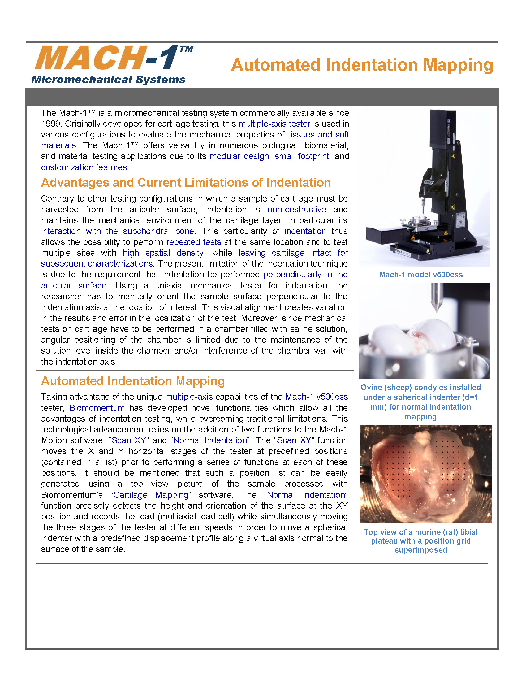
Articular surfaces were attached to a testing chamber filled with PBS and equipped with a camera-registration system.
-
A position grid was superimposed on the image of the sample (Fig. 1).
-
Pixels were converted into metric coordinates for the automated surface mapping.
Automated Indentation
At each position, a perpendicular indentation was performed by simultaneously moving the 3 displacement components (motorized stages) (Fig.2) based on the surface orientation and the resulting force is measured with a spherical indenter (r=0.5mm) placed on a multiaxial load cell.
Automated Thickness Measurement
-
The spherical indenter was replaced with a Needle probe (26G).
-
The thickness was obtained by penetrating the cartilage surface down to the subchondral interface.
-
The Cartilage surface corresponded to the position where the force started to increase, the subchondral interface where the force increased steeply and the vertical thickness was the difference. (Fig.3)
Thickness = Vertical thickness x cosine (surface orientation)
Extraction of the Instantaneous Modulus
-
The Instantaneous Modulus at each position was obtained by fitting the load-displacement curve to an elastic model in indentation using the measured thickness. (Fig. 4)
Results
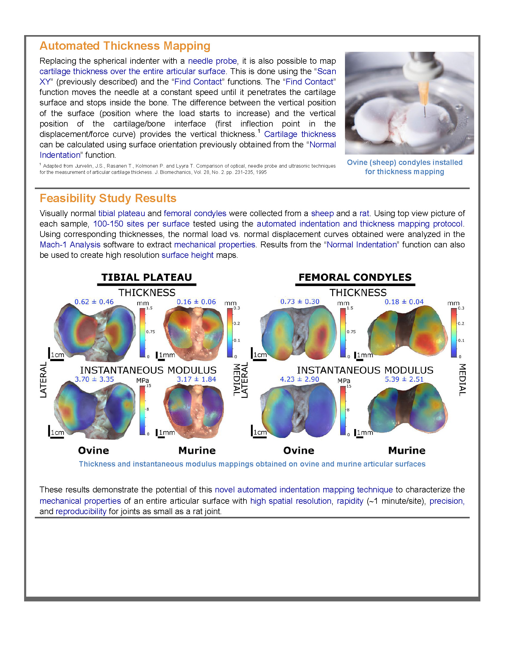
Spatial distribution of thickness and instantaneous modulus (Fig. 5) reveals a large variation within the medial and lateral projections of the femoral condyle and tibial plateau. This trend can be observed for all three species. The cartilage is thinner and stiffer in regions covered by the meniscus while a thicker and softer cartilage is observed over the rest of the surface.
Measured thickness agrees with those reported in the literature. The measured instantaneous modulus mappings show similar distribution patterns than those previously observed for the stifle joints of larger species, with stiffer cartilage in the region covered by the meniscus, suggesting a dependence with weight bearing and kinematics.
Conclusion
Cartilage thickness and instantaneous modulus can vary by a factor up to 10 over a distance of only 5% of the total articular surface width. These thickness and modulus maps clearly show that any difference between treated and non-treated cartilage could be confounded with the natural topographic variability rather than due to the treatment itself. By considering the spatial distribution of cartilage properties when choosing control and treated sites, the effects of treatment may be more easily discerned.
RELEVANCE OF THE SPATIAL DISTRIBUTION OF MECHANICAL PROPERTIES OF ARTICULAR CARTILAGE IN ANIMAL STUDIES
In order to evaluate the effect of cartilage treatments, appropriate control sites need to be chosen. However, finding the right location for the control site is quite challenging in articular surfaces where a natural spatial distribution of mechanical properties is present in all healthy joints. The purpose of this study was to assess the importance of considering the spatial distribution of the mechanical properties of normal articular cartilage in animal models of cartilage repair, specifically the distributions of thickness and instantaneous modulus.
Samples
Samples
- letally mature animals
- Right and left joints
-
Visually normal articular surfaces:
- Tibial plateau
- Femoral condyles
Camera-registration system
- Articular surfaces were attached to a testing chamber filled with PBS and equipped with a camera-registration system.
-
A position grid was superimposed on the image of the sample (Fig. 1).
- Pixels were converted into metric coordinates for the automated surface mapping.
Automated Indentation
At each position, a perpendicular indentation was performed by simultaneously moving the 3 displacement components (motorized stages) (Fig.2) based on the surface orientation and the resulting force is measured with a spherical indenter (r=0.5mm) placed on a multiaxial load cell.
Automated Thickness Measurement
- The spherical indenter was replaced with a Needle probe (26G).
- The thickness was obtained by penetrating the cartilage surface down to the subchondral interface.
-
The Cartilage surface corresponded to the position where the force started to increase, the subchondral interface where the force increased steeply and the vertical thickness was the difference. (Fig.3)
Thickness = Vertical thickness x cosine (surface orientation)
Extraction of the Instantaneous Modulus
Spatial distribution of thickness and instantaneous modulus (Fig. 5) reveals a large variation within the medial and lateral projections of the femoral condyle and tibial plateau. This trend can be observed for all three species. The cartilage is thinner and stiffer in regions covered by the meniscus while a thicker and softer cartilage is observed over the rest of the surface.
Measured thickness agrees with those reported in the literature. The measured instantaneous modulus mappings show similar distribution patterns than those previously observed for the stifle joints of larger species, with stiffer cartilage in the region covered by the meniscus, suggesting a dependence with weight bearing and kinematics.
- The Instantaneous Modulus at each position was obtained by fitting the load-displacement curve to an elastic model in indentation using the measured thickness. (Fig. 4)
Spatial distribution of thickness and instantaneous modulus (Fig. 5) reveals a large variation within the medial and lateral projections of the femoral condyle and tibial plateau. This trend can be observed for all three species. The cartilage is thinner and stiffer in regions covered by the meniscus while a thicker and softer cartilage is observed over the rest of the surface.
Measured thickness agrees with those reported in the literature. The measured instantaneous modulus mappings show similar distribution patterns than those previously observed for the stifle joints of larger species, with stiffer cartilage in the region covered by the meniscus, suggesting a dependence with weight bearing and kinematics.
Conclusion
Cartilage thickness and instantaneous modulus can vary by a factor up to 10 over a distance of only 5% of the total articular surface width. These thickness and modulus maps clearly show that any difference between treated and non-treated cartilage could be confounded with the natural topographic variability rather than due to the treatment itself. By considering the spatial distribution of cartilage properties when choosing control and treated sites, the effects of treatment may be more easily discerned.