- Product Detail
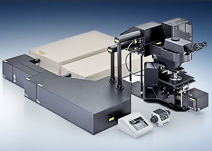
The Olympus FVMPE-RS multiphoton imaging system is purpose-built for deep imaging in biological tissue, aimed at revealing both detail and dynamics. Innovative features for efficient delivery and detection of photons in scattering media enable high signal-to-noise ratio acquisition. This translates to bright images with precise details — even from deep within the specimen. High sensitivity is matched with high-speed imaging to capture rapid in vivo responses. For advanced applications, dual-wavelength excitation extending to 1300 nm is available. Independent control of visible or multiphoton laser light stimulation and the ability to synchronize with patch clamp data are also possible.
Multiple configurations are available on the FVMPE-RS platform; each imaging system is customizable to meet your unique research requirements.
Designed for Deep Imaging
Maximize Resolution in Deep Imaging
Olympus TruResolution objectives maximize resolution and contrast for 3D imaging deep within thick specimens. The objectives are equipped with a motorized correction collar that can automatically and dynamically compensate for spherical aberration while maintaining focus position.
Refractive index mismatch between the sample and immersion medium introduces spherical aberration that degrades the focus of an objective lens. This aberration worsens deeper into the sample, yielding progressively poorer images with depth. TruResolution objectives solve this challenge with an autocorrection collar. The system can be driven either by an algorithm that finds the best collar setting at each depth or by user-defined manual presets to deliver consistently bright and sharp images through out the captured volume.
Maximize Signal with Deep Focus Mode
Deep focus mode adjusts the diameter of the laser beam based on the laser scattering conditions. For in vivo specimens with heavy laser scattering, the beam is narrowed so that more excitation photons reach deeper within your sample, helping produce bright, high-resolution images.


Laser with Negative Chirp Improves Excitation Efficiency at the Focal Plane
A laser beam with optimally adjusted pulse widths can be delivered to the focal plane, thanks to the application of negative dispersion that perfectly corresponds to the magnitude of the pulse-width dispersion generated during transmission through the microscope optics. The result is brighter images without needing to increase laser power, sample heating, photobleaching and phototoxicity.

Observe In Vivo and Cleared Tissue Specimens Up to 8 mm Deep
Olympus dedicated multiphoton objectives are optimized for deep imaging under in vivo and cleared tissue conditions. A diverse lineup provides you the opportunity to select an objective according to your research requirements. Different optical designs emphasize high numerical aperture, long working distance, wide field of view, and compatibility with a range of immersion media and tissue clearing agents.


Silicone Oil Immersion for Live Cell Imaging
The UPLSAPO30XSIR objective applies the efficient IR transmission coating of MPE dedicated objectives to a high numerical aperture silicone oil immersion objective. This combination makes the objective well-suited to multiphoton imaging of live cell specimens. The closer refractive index matching between silicone oil and live cells minimizes image distortion in the Z-direction and improves image brightness and resolution.
During time-lapse imaging, the refractive index of silicone oil remains constant, and the oil does not dry out, minimizing the amount of time researchers need to spend tending the experiment.


Visible to IR Transmission with Olympus 1600 Coating
Olympus multiphoton objectives and scanner optics have an optical coating that offers excellent transmission from 400 nm to 1600 nm. Efficient infrared transmission translates to more available power for fluorescence excitation at depth while strong support at short wavelengths maintains efficient collection of fluorescence and harmonic emissions.

Capture Fast, Dynamic Cellular Processes
High-Speed, 438 Frames-per-Second Scanning
A fast resonant scanner and conventional galvanometer scanner provide high-speed and high-resolution imaging in a single system. Avoid motion artifacts when imaging dynamic samples with capture rates of 30 fps at 512 × 512 pixels at the full field of view (FN 18) or up to 438 fps at 512 × 32 pixels. These speeds enable applications such as tracking of fast moving cells in blood flow, and observing rapid membrane potential dynamics across neurons and other cells.


Efficient Laser Transmission
Olympus silver-coated scanner mirrors help deliver more laser power to your sample to better excite fluorescence, and yield brighter images. The silver-coated mirrors achieve very high re-flectance across a broad wavelength range, from visible to near infrared. The total reflectance for the XY scanner is particularly improved in the near infrared range compared to conventional aluminum-coated mirrors. The increased reflectance helps maximize available laser power and deliver the power needed for deep in vivo experiments.

A Cooled, High-Sensitivity GaAsP Detector Acquires High S/N Images
High signal-to-noise ratio imaging can be acquired even from faint fluorescence through the use of high sensitivity gallium arsenide phosphide (GaAsP) photomultiplier tube (PMT) detectors. — GaAsP PMTs deliver greater quantum efficiency than standard multialkali PMTs. Fan-less Peltier cooling further improves the signal-to-noise ratio. You can also leverage the advantages of both detector types by combining them in a single system.


Microsecond Timing for Electrophysiology and Optogenetics
A hardware sequencer provides microsecond precision timing for stimulation and triggering events. Stimulation can be spatially and temporally synchronized to the imaging scan, facilitating the capture of fast response dynamics at precise locations. In the context of electrophysiology and optogenetics, this could mean the difference between distinguishing a synchronous versus an asynchronous stimulus response. For acquisitions lasting two weeks or longer or experiments with complex procedures that require switching between imaging tasks, the sequence manager software module maintains millisecond precision, providing high-quality data in demanding in vivo and in vitro experiments.

Spend Less Time Tuning the Laser
Multiwavelength Excitation for Multiphoton Imaging
The FVMPE-RS imaging platform supports a dual wavelength infrared pulsed laser or two independent infrared lasers for multichannel, multiphoton excitation imaging. You can optimally excite different fluorophores without having to repeatedly tune the laser. Simultaneous excitation with independent power control of each laser line enables users to capture balanced images of different fluorophores. Separate excitation wavelengths for individual fluorophores can also reduce background tissue autofluorescence by shifting excitation away from the 800 nm range.

Efficient Longer Wavelength Excitation
The InSight X3 pulsed IR laser by Spectra-Physics supports multiphoton imaging with tunable excitation from 680–1300 nm. The dual-line version adds a second fixed line at 1045 nm. With improved laser power beyond 1000 nm, the InSight X3 laser provides access to new multiphoton imaging capabilities. Utilize the growing library of red-shifted dyes and fluorescence proteins for deeper imaging or broader multichannel coverage. Perform third harmonic generation imaging in biological specimens without UV damage.


User-Friendly, Accurate Imaging with Auto Laser Alignment
Quadralign 4 axis laser alignment simplifies system upkeep by maintaining the precise alignment of the excitation beam into the scanner unit, even in the face of laser drift due to wavelength tuning, temperature fluctuation, and other sources of cavity shift. The beam position and angle are automatically adjusted to deliver higher laser power and consistent pixel registration. If your system has two excitation laser lines, this feature offers an additional benefit. Auto laser alignment maintains co-alignment between the beams, helping eliminate co-registration errors between channels. The laser alignment can also be manually fine-tuned using the software interface.

Optional Features for Advanced Applications
Simultaneous Multiphoton Stimulation and Imaging with the SIM Scanner
The SIM scanner, an independent galvanometer scanner, and visible laser modules can be added for precise microsecond photostimulation and photobleaching experiments. On systems with two IR imaging lines, the SIM scanner enables simultaneous multiphoton stimulation and imaging.

Wide Choice of Scan Modes
The FVMPE-RS comes with AOM as standard and provides fine position and time control of imaging and light stimulation. Using Olympus’ own tornado scanning allows rapid bleaching and laser light stimulation of desired fields in experiments.

Synchronize Electrophysiological Data and Laser Light Stimulation with the Analog Unit
Analog inputs and TTL I/O are available to support electrophysiology experiments. The analog input unit records external voltage signals as images that are treated the same way as normal image data. Light-stimulated electrical signals measured with patch clamps can be synchronized with image capture and displayed as a pseudocolor intensity overlay.


3D Multi Point Measurement Provide Rapid Intensity Measurement with High Signal to Noise
Raster scanning takes time. With MMASW(Multi-Point Mapping Software) multi-point acquisitions you can eliminate that time by placing the laser only where needed, retaining high signal to noise output and allowing you the freedom to choose and optimize your scan path. A scan of multiple positions can go as fast as 101 Hz, and each position can gather signals at up to 50,000 Hz, providing you with highly relevant physiological data.

Multi-point mapping software is designed for extremely fast functional measurements in living cells or tissues where researchers use light to probe fluorescent intensity changes. Resonance scanners or acousto-optic deflectors (AODs) are intrinsically less sensitive due to their very short integration times and the number of photons that can be detected. By retaining high integration time per position where it is needed in a single point laser scan, the multi-point mode allows greater multiphoton depth penetration and signal-to-noise. Each point can also be expanded to an array for larger area stimulation or detection.
Synchronize your measurement scans with simultaneous stimulation using the SIM scanner and free your functional imaging.
Create 3D Stimulation Reaction Maps
To achieve highly targeted laser light stimulation, the observation field is divided into a grid, and the laser illuminates each area in a pseudorandom sequence that avoids sequential stimulation of adjacent areas. A stimulation reaction map is drawn based on patch clamp recording or imaging intensity. Integration of an optional piezo nosepiece extends the reaction map to 3D, with stimulation delivered at depths different from the imaging plane.


Intuitive Software Optimized for Multiphoton Observation
Customizable Layout
Customize which controls you see in the software interface and where they are located. Save and reload your favorite layouts.

Save and Recall Settings
Save the acquisition parameters that you use during an experiment. The parameters can be easily recalled for repeatable imaging conditions.
Precisely Control Your Experiments
The sequence manager makes it easy to coordinate experiments. Complex protocols, such as changing the frame rate during time-lapse imaging or repeating photostimulation events at different positions during image acquisition, can be organized and accurately carried out with precise timing. Protocols can also be saved and later reloaded for consistent execution of experiments.

Combine Wide Field of View with High Resolution
See your entire specimen at high resolution and in context with the tiling function. This software feature scans multiple adjacent images and stitches them together. With a motorized stage, images can be stitched together in a very wide field of view, while the mapping feature makes it easy to locate the position of specific cells in the larger image.

Expanded Analysis Functions
The FVMPE-RS imaging platform’s software is integrated with Olympus cellSens image analysis software, expanding the system’s analytical capabilities. Optional features include 3D deconvolution for Z-stack images, area estimation for each particle in an image, an image processing filter, and colocalization analysis.
Separate Overlapping Channels with Spectral Deconvolution
Closely overlapping fluorescence spectra can complicate biological studies that look at multiple labels simultaneously. Separation of overlapping spectral channels is possible via spectral deconvolution based on either a blind unmixing algorithm or previously saved multichannel profiles. Cross-talk between the channels can even be eliminated during image acquisition via live processing.


3D Rendering
Large amounts of Z-stack data can be rendered into a 3D display. Important views can be registered as key frames, making it easy to create animated views of 3D images that zoom and transition to different camera angles.

Choose From 3 Frames and 3 Laser Configurations
Microscope Frames
Upright Microscope System — Designed for Multiphoton Microscopy

This Upright Frame is completely dedicated to multiphoton microscopy. Providing space for large samples, a high degree of motorization and nosepiece focus control enables the stage and your sample to remain fixed and stable.
Gantry Microscope System — For in vivo Observation that Require Maximum Space

With its ultra-stable arch-like structure, the new Gantry Frame offers tremendous space beneath the objective lens along with a high degree of flexibility to suit different samples. This is ideal for in vivo observation requiring maximum space.
Inverted Microscope System — For Observing Tissue Cultures, 3D Cultures, and Cell Cultures (Spheroid)

The Inverted Frame is ideal for the time lapse observation of thick, living specimens such as tissue cultures, and three-dimensional cell cultures.as The inverted frame system also finds utility in intravital time lapse observation of organs and tissues through a body window.
Laser Systems
One Laser System

This streamlined system uses a single multiphoton infrared laser for imaging. Add an optional SIM scanner for visible laser stimulation.
Dual Laser Lines System

This system supports dual wavelengths for multiphoton, multicolor imaging. Add an optional SIM scanner for visible laser stimulation and simultaneous multiphoton stimulation at 1045 nm.
Twin Lasers System

This flexible system employs two multiphoton infrared lasers for imaging. In addition to multiphoton, multicolor imaging, visible laser stimulation and simultaneous multiphoton stimulation across tunable wavelengths is also supported in combination with an optional SIM scanner.
Lasers for Multiphoton Configurations
The InSight X3 Dual-OL laser enables dual wavelength, simultaneous imaging for deep observation. It has a high peak power with short, 120 fs pulse widths, a broad continuously tunable range from 680 nm to 1300 nm, and a fixed wavelength line at 1045 nm. A selection of other models are available to match your multiphoton excitation requirements.
Visible Beam Combiner for Laser Light Stimulation

The laser combiner enables solid-state laser combinations for laser light stimulation at wavelengths of 405 nm, 458 nm, and 588 nm.
Modular Units Designed for Your Applications
Light Guide Illumination
Light Guide Illumination Source U-HGLGPS

This light source is equipped with a liquid light guide that minimizes the impact of vibration and lamp heat on both the microscope and specimens. With a metal-halide bulb, the light source offers an average lifetime of 2000 hours.
Non-descanned Detectors
Transmitted Non-descanned Light Detector

A high NA condenser and transmitted non-descanned light detector for multiphoton imaging detect fluorescence and harmonic emissions scattered in the forward direction from the sample plane.
Reflected Non-descanned Light Detector
Multialkali PMT 2CH Detector

This basic multialkali 2CH PMT provides robust performance across a wide range of wavelengths.
Multialkali PMT 4CH Detector

Expand your simultaneous detection capability with a total of 4 multialkali PMTs.
Multialkali PMT 2CH + 2CH Cooled GaAsP PMT Detector

Combine the robustness and dynamic range of multialkali PMTs with the high sensitivity of GaAsP PMTs.
Cooled GaAsP PMT 2CH Detector
This Peltier-cooled 2CH GaAsP PMT provides higher sensitivity for weak signals or short pixel dwell times.
-
Olympus is one of the world’s leading manufacturers of professional opto-digital products for medicine, science and industry. As a result, Olympus provides a comprehensive range of solutions. From microscopes for training and routine tasks to high-end system solutions in the fields of life science, there is a system for every need. The product line is complemented by innovative laboratory equipment for cellular research applications and the new all-in-one microscopes that offer user engagement at all levels.
| Request Infomation |
| Related News |
| Recently viewed products |
