|
1. SynBBB 3D Blood Brain Barrier Model – Real-time visualization of cellular and barrier functionality SynVivo’s SynBBB 3D blood brain barrier model recreates the in vivo microenvironment by emulating a histological slice of brain tissue cells in communication with endothelial cells across the blood brain barrier (BBB). Shear-induced endothelial cell tight junctions, which cannot be achieved in the Transwell® model, are easily achieved in the SynBBB model using physiological fluid flow. Formation of tight changes can be measured using biochemical or electrical analysis (assessing changes in electrical resistance) with the SynVivo Cell Impedance Analyzer. Interactions between brain tissue cells and endothelial cells are readily visualized in the SynBBB assay. Transwell models do not allow real-time visualization of these cellular interactions, which are critical for understanding of the BBB microenvironment. SynBBB is the only in vitro BBB model that allows: ●Accurate in vivo hemodynamic shear stress ●Real-time visualization of cellular and barrier functionality ●Significant reduction in cost and time ●Robust and easy to use protocols  The SynBBB system is a highly versatile platform for investigation of: Tight junction proteins: Determine the levels of tight junction proteins namely zonula occludens, claudins and occludins which regulate the BBB. Transporter proteins: Analyze functionality of transporter proteins (e.g. Pgp) in normal and dysfunctional BBB. Drug permeability: Evaluate real-time permeability of therapeutics and small molecules across the endothelium of the BBB. Inflammation: Understand the underlying mechanisms of inflammatory responses on the regulation of the BBB. Cell migration: Visualize and quantify in real-time migration of immune cells across the BBB. Omic changes: Perform genomic, proteomic and metabolic analysis on normal and dysfunctional BBB. Neurotoxicity: Analyze toxicity effects of chemical, biological and physical agents on the cells of the BBB. Neuro-oncology: Investigate effects of the tumor cells on the BBB. Depending on your research needs you can select from the “basic” SynBBB model or a “TEER-compatible” SynBBB configuration. 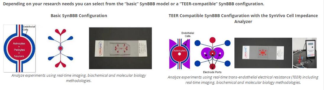
TEER Compatible SynBBB Configuration with the SynVivo Cell Impedance Analyzer Analyze experiments using real-time trans-endothelial electrical resistance (TEER) including real-time imaging, biochemical and molecular biology methodologies. | |
|
2. SynTumor 3D Cancer Model – Recreating the tumor microenvironment SynTumor is a 3D tissue model for real-time visualization and quantitative assessment of cell-cell and cell-drug interactions in a physiologically and morphologically realistic tumor microenvironment. The system enables (a) circulation in the microvasculature, (b) transport across the vessel walls, and (c) delivery to the tumors. Starting with scans of vascular networks incorporated with interstitial and tissue/tumor spaces, the SynTumor 3D tissue model creates an in-vitro tumor microenvironment akin to a viable histological slice. The SynTumor 3D Cancer model has the following benefits: ●Side by side architecture enables quantitative real time visualization ●Physiological leaky vasculature with engineered porous structures ●Morphologically realistic in vivo based architecture ●Physiologically realistic convective and diffusive transport ●Microfluidic platform with ultra-low consumable volumes Depending on your research needs you can select from the “idealized” or “microvascular” SynTumor 3D Cancer Model configuration. 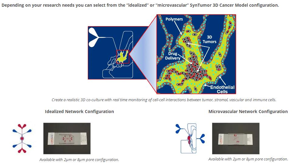
| |
|
3. SynRAM 3D Inflammation Model – Visualize Rolling, Adhesion and Migration in a Single Assay The SynRAM™ 3D Inflammation Model from SynVivo has been developed to study the entire inflammation pathway in a realistic and dynamic environment. By recreating a histological slice of co-cultured tissue and/or tumor cells with a lumen of endothelial cells, the SynVivo platform delivers a physiologically realistic model including flow and shear in a platform and enables real-time tracking of rolling, adhesion and migration processes. This model has been successfully validated against in vivo studies showing excellent correlation with rolling velocities, adhesion patterns and migratory processes (Lamberti et al 2014, Soroush et al 2016). 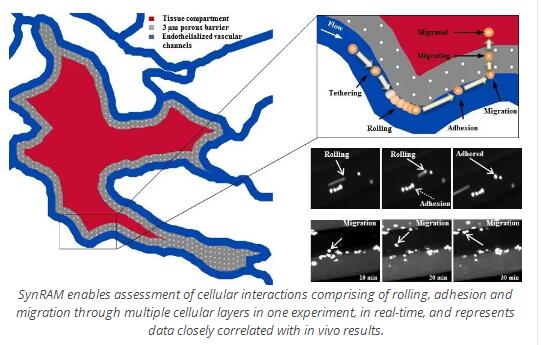 The SynRAM 3D inflammation model provides a realistic testing environment including: ●Physiological shear stress within a microvascular environment ●In vivo like vascular morphology with fully enclosed lumen ●Co-culture capability for cell-cell interactions ●Quantitative real-time rolling, adhesion, and migration data from a single experiment SynRAM enables assessment of cellular interactions comprising of rolling, adhesion and migration through multiple cellular layers in one experiment, in real-time, and represents data closely correlated with in vivo results. SynRAM’s innovative design overcomes the current limitations inherent in flow chambers or Transwell chamber based assays. Current flow chamber designs are oversimplified, lack the scale and geometry of the microenvironment and cannot model transmigration. Similarly, Transwell chambers do not account for fluid shear and size/topology observed in vivo, the end point measurements of migration are not reproducible and do not provide real-time visualization. SynVivo’s proprietary chip designs range from complex in vivo derived microvascular networks (obtained from digitized images) to produce realistic cellular makeup and vascular morphology resulting in varying shear and flow conditions, to simplified idealized networks designed to reproduce the cellular makeup 
| |
|
4. SynTox 3D Toxicology Model – Organ Specific Physiological Responses SynTox is the only commercially available 3D toxicology model with real-time optical monitoring and multi-compartment, multi-cellular architecture and low reagent requirements. Other benefits of this platform are: Physiologically realistic morphological, fluidic and 3D cellular conditions Universal platform with architecture specific of desired organ Significant reduction in cost and time Robust and easy to use protocols Compatible with standard analytical instruments for both on chip and off chip assays including omic methodologies for systems biology and bioinformatic analysis SynTox 3D Toxicology Model recreates the in vivo micro-environment by emulating a histological slice operating in an in vitro Environment. 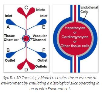 Current in vitro platforms are poor predictors of in vivo safety, efficacy and pharmacokinetics of therapeutics owing to significant difference in the test conditions compared to physiological conditions observed in vivo.
 Current in vitro models routinely utilize 2D monolayers or 3D aggregates of cells under static conditions for studying drug toxicity. These models fail to reproduce in vivo physiological features such as morphological size, physiological blood flow and cellular (biological) make-up of the specific organs being investigated. Other microfluidic models employ a membrane-based top-bottom two-compartment architecture, inherently limiting key desired features such as real-time visualization and the ability to simultaneous analyze multi-cellular cultures.
| |
 单细胞应力加载系统,单个细胞机械力刺激培养系统
|
 单层细胞静水压力培养系统
|
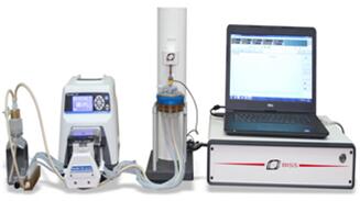
 加拿大 多功能组织材料微观生物力学测试分析系统
|
 加拿大 软骨质量评估分析系统
|
 提供瑞士regenhu品牌3D生物打机样机、生物打印科研委托服务,申请电话:010-67529703
|
 CellSorter自动、无损伤、高精度、高通量单细胞捕获分选分析系统
|
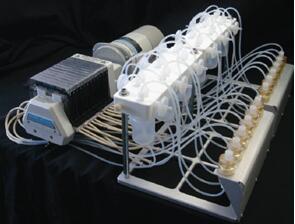
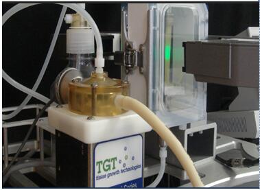
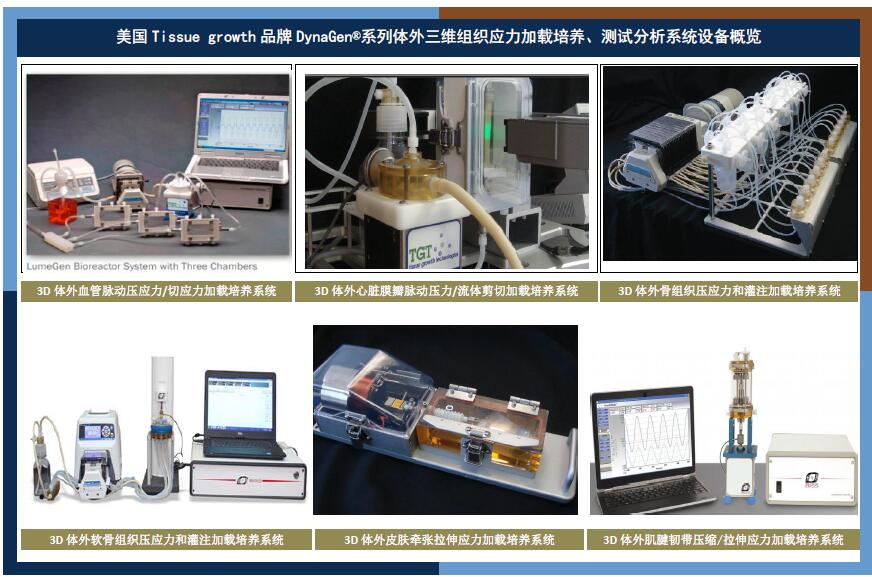 美国tissue growth三维血管、软骨、肌腱韧带、皮肤生物反应器美国tissue growth三维血管、软骨、肌腱韧带、皮肤生物反应器
|
 流式单细胞力学特性测试实时分析和形变实时测量系统
|
 欧美进口细胞牵引力显微镜和微柱
|
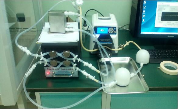
 加拿大cellscale生物组织材料机械力特性测试分析和细胞组织刺激仪系列国代理
|
- Mechanobiology
- 3D BioPinter
- Cell analysis&Cell Sort
-
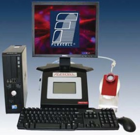
flexer-cell® FX-6000™ Tension System (New)
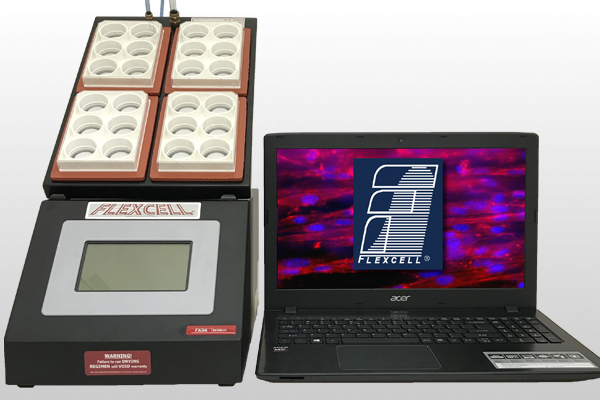
Feature:Apply equibiaxial or uniaxial tension to cells in 2D or 3D culture,allow the user to observe signaling responses to strain in real time on a microscope stage.
Uses vacuum pressure and positive air pressure to deform a flexible-bottomed culture plate yielding up to 33% substrate elongation. Mimics in vivo conditions in cells from muscle, lung, heart, vascular vessels, skin, tendon, ligament, cartilage, and bone. Applies a defined, controlled, static or cyclic deformation to cells growing in vitro. Control of the vacuum pump with the FX-6000™ software. Compatible with Windows 10.flexer-cell Jr. Tension system
Feature:Apply equibiaxial or uniaxial tension to cells in microscope devices,use flexer-cell® StageFlexer® Jr. A compact strain device designed to accept membranes removed from the well of a BioFle®x®, Tissue Train®, or UniFlex® culture plate and observe signaling responses to strain in real time.flexer-cell cell/tissue Compression System
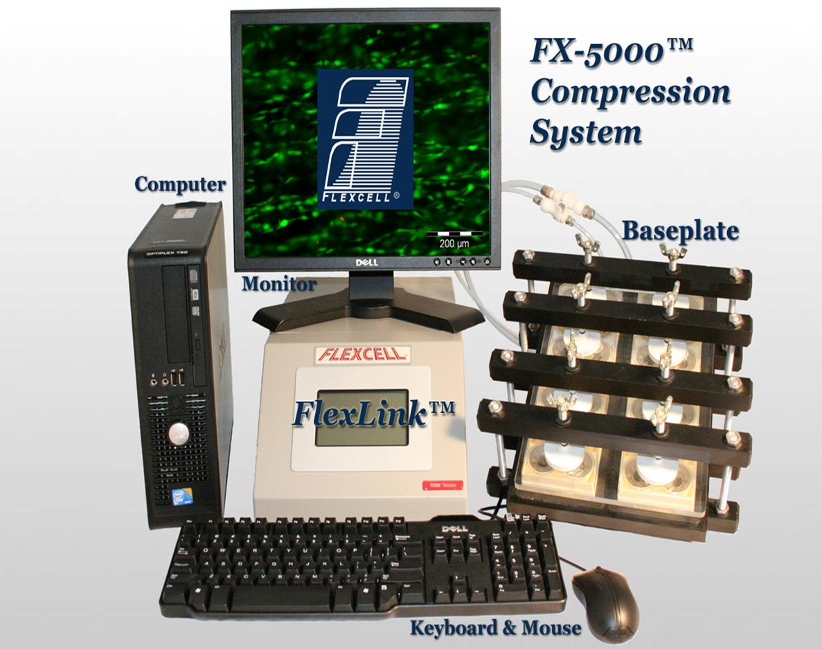
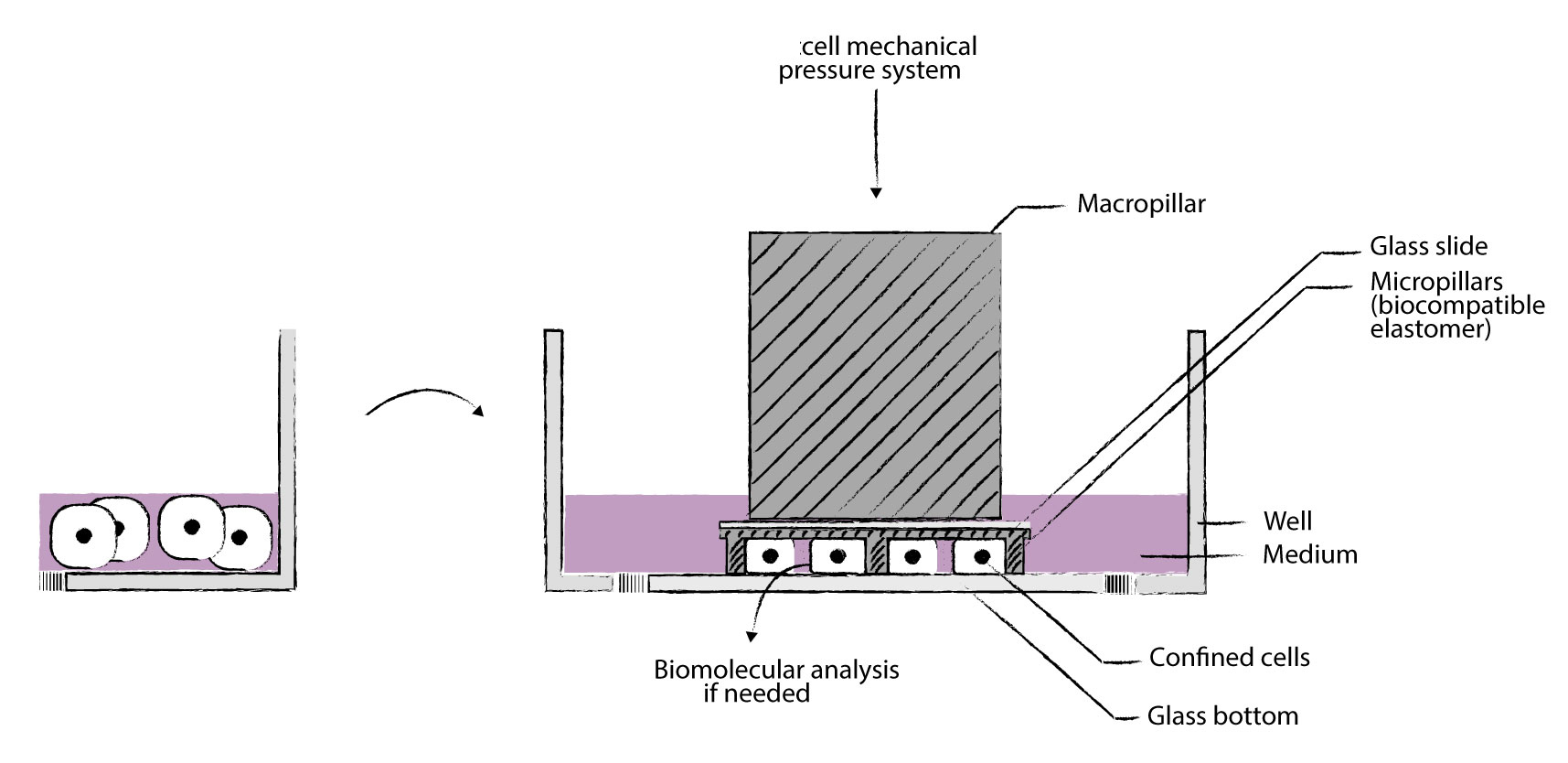
Feature:Apply compression to tissue samples or 3-D cell-seeded constructs, use flexer-cellFeature StagePresser™--A single-well embodiment of the compression apparatus designed to allow the user to detect signaling responses to compression.flexer-cell 3D cell and 3D Tissue Train System
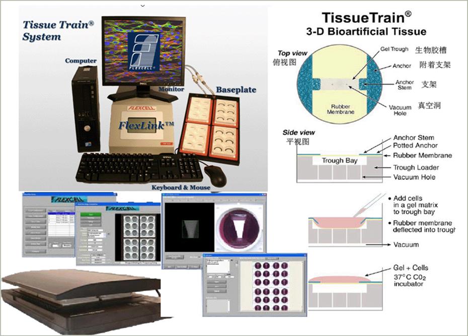
Feature:3-D cell culture in a gel matrix with or without cyclic uniaxial tension,A computer-regulated bioreactor that allows users to create 3-dimensional cell-seeded gels and apply uniaxial tensile strains to these gels.flexer-cell® SCANFLEX™ WITH XYFLEX™
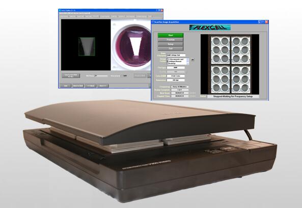
>Feature:Measure gel compaction in 3-D bioartificial tissues,Automated repetitive scanning process. Scans and saves images of 3-D tissue constructs.User defined frequency and time intervals of image capture. Can be used with the Tissue Train system to determine the compaction kinetics (change in area) of 3-D cell seeded-gels.Scans four 6-well Tissue train culture plates placed on the scanner bed. Images can be imported into Xyflex™ program to analyze area measurement.flexer-cell Streamer Fluid Shear System
Feature:Apply fluid shear stress to cells with laminar, pulsatile, or oscillating flow,use flexer-cell® FlexFlow™ Shear Stress Device--A shear stress device that allows the user to observe signaling responses to fluid flow and/or apply strain to cells before, during, or after applying a shear stress...
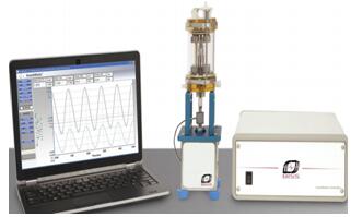
mach-1 micromechanical testing system
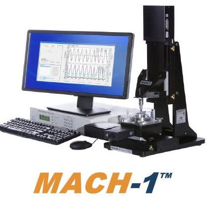
Mach-1™ is a micromechanical testing system commercially available since 1999. Originally developed for cartilage testing, this multiple-axis tester is used in various configurations to evaluate the mechanical properties of tissues and soft materials. Due to its modular design, small footprint and customization features, the Mach-1™ offers versatility in numerous biological, biomaterial, and material testing applications.
Arthro-BST™ device
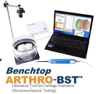
The hand-held Arthro-BST™ device is used to map electromechanical properties of articular cartilage. The purpose of the study was to evaluate correlation of electromechanical properties with histological, biochemical and biomechanical properties of cartilage& 3D Vascular Bioreacto

The DynaGen® Series bioreactors provide mechanical stimulation to three dimensional tissue engineered constructs to create physiological conditions in-vitro. The system imparts pulsatile flow and pressure to a vascular conduit. Applications include the investigation of cell function and differentiation, pharmaceutical benchtop testing, and the seeding and growth of engineered tissues and medical devices& HeartValve Bioreactor

The DynaGen® Series bioreactors provide mechanical stimulation to three dimensional tissue engineered constructs to create physiological conditions in-vitro. The system imparts pulsatile flow and pressure to a valve or valve-like structure. Applications include the investigation of cell function and differentiation, pharmaceutical benchtop testing, and the seeding and growth of engineered tissues and medical devices.& Ligament and Tendon Bioreactor

he DynaGen® Series bioreactors provide mechanical stimulation to three dimensional tissue engineered constructs to create physiological conditions in-vitro. The system imparts axial stress or strain to a ligament or tendon construct. The stress/strain profiles can be defined by the user and can be a simple harmonic (sinusoidal) or a physiological waveform. Applications include the investigation of cell function and differentiation, pharmaceutical benchtop testing, and the seeding and growth of engineered tissues and medical devices. Researchers are currently utilizing these systems in a wide range of research areas including:1)ACL regenerative medicine.2)Collagen gel stimulation.3)Small diameter tendon stimulation Hand tendon and ligament regenerative medicine& OsteoGen Bioreactor
's OsteoGen bioreactors impart perfusion through cell seeded cylindrical scaffolds. Applications include investigating cell function, modulating the growth and development of engineered tissues, or acting as a test bed for drug and regenerative medicine technologies. Researchers are currently utilizing these systems in a wide range of research areas including:1)Bone stem cell phenotype research2)Mineral deposition of marrowstromal cells& Cartilage Bioreactor

The DynaGen® Series bioreactors provide mechanical stimulation to three dimensional tissue engineered constructs to create physiological conditions in-vitro. The system imparts compressive or hydrostatic stress to a cartilage construct. The stress/strain profiles can be defined by the user and can be a simple harmonic (sinusoidal) or a physiological waveform. Applications include the investigation of cell function and differentiation, pharmaceutical benchtop testing, and the seeding and growth of engineered tissues and medical devices. Researchers are currently utilizing these systems in a wide range of research areas including:1)Stem cell stimulation. 2)Meniscus regenerative medicine.3)Chondrocyte stimulation& 3D skin Bioreactor

The bioreactors impart static and oscillatory stimulation to a "skin like" sample in a unique air-to-nutrient media interface. Applications include investigating cell function and stimulating the growth and development of engineered tissues. The device can also be used as a "test bed" for pharmaceutical and regenerative therapies. The can apply either stress or strain using operator defined wave forms -
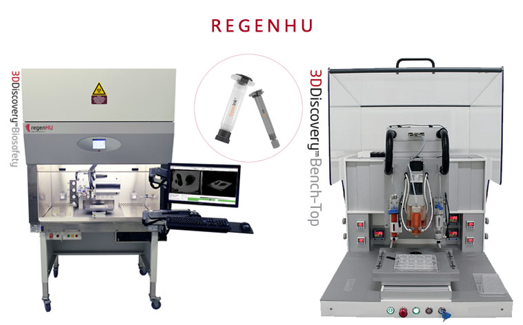
RegenHU 3Ddiscovery 3D bio-printing platform

The 3DDiscovery® instrument is a cost-effective 3D bio-printing platform to explore the potential of 3D tissue engineering through the bio-printing approach.
RegenHU BioFactory® high end, versatile and cell friendly three dimensional bio-manufacturing instrument
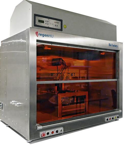
The BioFactory® is a high end, versatile and cell friendly three dimensional bio-manufacturing instrument. It allows researchers to pattern cells, biomolecules and a range of soft and rigid materials in desirable 3D composite structures in order to mimic biomimetic tissue models.
-
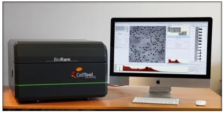
BioRam®System— Live cells Raman spectroscopy system
Features:
Photonic fingerprinting® for gentle cell analysis,Raman Trapping Microscopy for stress- and label-free cell identification and characterization
1)Identifying cells from their biophotonic profile
Raman spectroscopy is based on the Raman effect: when a molecule is exposed to laser light, a small fraction is scattered with a shift in frequency compared to the incident light. This shift is highly specific for each molecule – as unique as a fingerprint. Raman spectroscopy has long been used successfully as a contact-free technique for material characterization. More recently, scientists have demonstrated its applicability to the field of biology and medicine: several research teams have independently shown that the combined Raman spectra of all biopolymers within a cell form distinct clusters according to cell type and state of differentiation and activity
2)Mastering the challenges of biological samples
In close collaboration with scientists at the Fraunhofer Institute for Interfacial Engineering and Biotechnology, CellTool have overcome the particular technical challenges that have so far hampered routine use of Raman spectroscopy in biomedical research and development. Biological samples often emit fluorescence which interferes with their weak Raman signals. Furthermore, they require a tolerable wavelength and appropriate physiological conditions during measurement to preserve cell viability.
3)Raman spectroscopy for living cells
BioRam, CellTool’s most advanced analytical platform, is available for customers.
4)The measurement process: Just get going
Samples can be used without special preparation: as living tissue, in aqueous solution on Raman-compatible slides or directly in culture medium in Raman-compatible petri dishes or fluidic chips. With a 3D-mouse, you can home in on the cells to be analysed and start the automatic generation of Raman spectra with a mouse click. Cells are then automatically identified according to their spectra by database comparison. Measurement coordinates, live images, raw data and all the relevant parameters are saved as well, enabling further analysis or the application of alternative algorithms at any time.
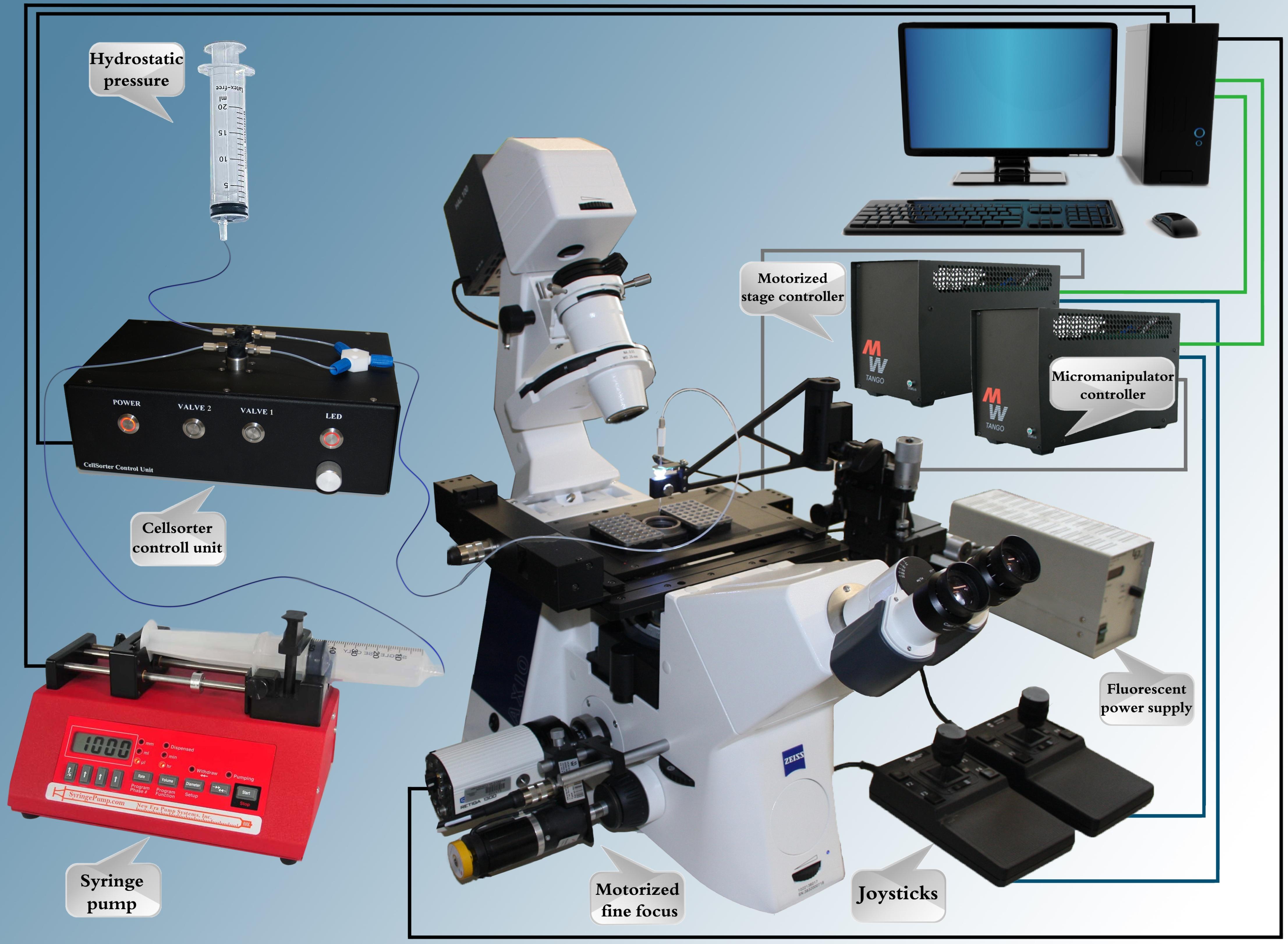
Cellsorter®Automated single cell sorting and deposition
Features:
How to get the one you want? Single cells are in your hands——sort live cells right from the petri dish
1)High throughput single cell sorting directly from the Petri dish
2)One single cell arrives to each PCR tube
3)10 PCR strips containing 80 tubes can be filled in a cycle
4)Glass cover slip for testing single cell deposition in situ
5)Drop volume less than 1 ul
6)15-20 seconds per cell
7)Isolates a subpopulation of live adherent cells expressing fluorescent or luminescent markers
8)Both unlabeled and fluorescent cells are recognized by computer vision
9)Viable cells after sorting
10)Sorts cells labeled by fluorescent molecular probes highlighting specific cellular activities
11)Collects single cells for further cultivation, cloning, RNA or protein preparation
12)Sorts fixed cells after immunocytochemical preparation
13)Any adherent cell type can be sorted
14)Cell culture needs minimal preparation before sorting
15)Average sorting process takes only a few minutes
16)Multichannel detection using the fluorescent filter setup of the microscope
17)When collecting multiple cells sorting speed is 1 cell/second. Number of cells picked up in a single run: 1-1000.calScreener™multi-channel calorimetric system for cellular

Features:
Monitor cell metabolism and cellular bioenergetics in real time with the label-free calScreener™ from SymCe
1)Monitor cell metabolism and cellular bioenergetics in real time
calScreener™ is the first multi-channel calorimetric system specifically developed for cell based assays. Measurement of heat flow in living cells is a well established and proven technology to monitor changes in metabolic processes. calScreener™ provides the researcher with a label-free detection tool with maximal versatility in assay type. The real time continuous measurement provides important kinetic data for interpretation of cellular mechanisms and responses to treatment. calScreener™ is suited to drug development and metabolic research providing a true phenotype response where the total status of the cell metabolism is monitored. calScreener™ is further suitable for toxicology tests, process development and environmental monitoring.
2)Mastering the challenges of biological samples
3)First Calorimeter designed for cell biological work
4)Label-free assay
5)Independent of cell morphology
6)Measures heat produced from cell metabolism
7)True PHENOTYPE response measured
8)Real-Time continuous measurement
9)Prior knowledge of pathways and specific target function not needed
10)Multiple compounds can be tested simultaneously for synergetic effects without knowledge of pathway interactions
11)Suitable for a wide range of applications
- Regenerative Medicine
- Electrospinning
- Optical(magnetic) tweezers
-
3D Vascular Bioreactor
Features:
The DynaGen® Series bioreactors provide mechanical stimulation to three dimensional tissue engineered constructs to create physiological conditions in-vitro. The system imparts pulsatile flow and pressure to a vascular conduit. Applications include the investigation of cell function and differentiation, pharmaceutical benchtop testing, and the seeding and growth of engineered tissues and medical devices
HeartValve Bioreactor
Features:
The DynaGen® Series bioreactors provide mechanical stimulation to three dimensional tissue engineered constructs to create physiological conditions in-vitro. The system imparts pulsatile flow and pressure to a valve or valve-like structure. Applications include the investigation of cell function and differentiation, pharmaceutical benchtop testing, and the seeding and growth of engineered tissues and medical devices.
Ligament and Tendon Bioreactor
Features:
The DynaGen® Series bioreactors provide mechanical stimulation to three dimensional tissue engineered constructs to create physiological conditions in-vitro. The system imparts axial stress or strain to a ligament or tendon construct. The stress/strain profiles can be defined by the user and can be a simple harmonic (sinusoidal) or a physiological waveform. Applications include the investigation of cell function and differentiation, pharmaceutical benchtop testing, and the seeding and growth of engineered tissues and medical devices. Researchers are currently utilizing these systems in a wide range of research areas including:1)ACL regenerative medicine.2)Collagen gel stimulation.3)Small diameter tendon stimulation Hand tendon and ligament regenerative medicine

OsteoGen Bioreactor
Features:
's OsteoGen bioreactors impart perfusion through cell seeded cylindrical scaffolds. Applications include investigating cell function, modulating the growth and development of engineered tissues, or acting as a test bed for drug and regenerative medicine technologies. Researchers are currently utilizing these systems in a wide range of research areas including:1)Bone stem cell phenotype research2)Mineral deposition of marrowstromal cells
Cartilage Bioreactor
Features:
The DynaGen® Series bioreactors provide mechanical stimulation to three dimensional tissue engineered constructs to create physiological conditions in-vitro. The system imparts compressive or hydrostatic stress to a cartilage construct. The stress/strain profiles can be defined by the user and can be a simple harmonic (sinusoidal) or a physiological waveform. Applications include the investigation of cell function and differentiation, pharmaceutical benchtop testing, and the seeding and growth of engineered tissues and medical devices. Researchers are currently utilizing these systems in a wide range of research areas including:1)Stem cell stimulation. 2)Meniscus regenerative medicine.3)Chondrocyte stimulation -
E-FIBER ELECTROSPINNING SYSTEM-STARTER KIT
Features:
E-Fiber electrospinning is a plug & Play setup for users who deal with electrospinning techniques for the first time or for expert users who intend to develop new materials and require a new and easy setup for their research.The Starter Kit, however simple and easy to use, still has all the requirements and strong points of the high-end versions, such as the Advanced and the XL models.

E-FIBER ELECTROSPINNING SYSTEM-DESKTOP BASIC VERSION
Features:
E-fiber electrospinning advanced version is optimal for research centers, companies and universities whose needs are to produce nanomaterials intended both for commercial and research purposes.
E-fiber is a fully-customisable electrospinning platform consisting of injection stages and application-specific collectors (static or movable), for the laboratory-scale manufacturing of polymeric-nanofibrous matrices. This device creates a powerful and highly versatile technique for the creation of various fibrous architectures with controlled fibre diameter and orientation. Its technology minimises influence on the electrospinning process. E-fiber offers various technological solutions to accurately control each process guaranteeing batch-to-batch reproducibility and precise control of fibre parameters such as diameter, orientation and texture. The equipment is user-friendly and safe thanks to the automatic safety devices that protect the operator.
E-FIBER ELECTROSPINNING SYSTEM-XL VERSION
Features:
The DynaGen® Series bioreactors provide mechanical stimulation to three dimensional tissue engineered constructs to create physiological conditions in-vitro. The system imparts pulsatile flow and pressure to a valve or valve-like structure. Applications include the investigation of cell function and differentiation, pharmaceutical benchtop testing, and the seeding and growth of engineered tissues and medical devices. -

Single Molecule Biophysics Magnetic Trap—Stretching and twisting single molecules
Features:
icoTwist is involved in nanomanipulation and single-molecule experiments in biology. Historically, single molecule experiments were among the first nanotechnologies, appearing in the 1990s to characterize biopolymers and proteins with unprecedented accuracy. The idea is that instead of analyzing the behavior of a great number of molecules as usually done in a test tube, you now analyze the behavior of a single molecule. Thus instead of measuring average behavior you have direct access to the biopolymer’s activity in real time.
Turnkey TIRF Microscopy complex
Features:
Turnkey TIRF Microscopy complex is compatible with Nikon, Olympus, Zeiss, and Leica
1)inverted microscopes. The complex includes:
2)Prism-based p-TIRFM flow system, and/or
3)Lightguide-based lg-TIRFM flow system, and/or
4)Objective-based o-TIRFM flow system
5)Multi-color computer-controlled illuminator
6)Low light EMCCD camera Andor iXon or similar
7)Optional digital fluidics SmartFlow
8)Optional temperature control system TC-40 – 25-40oC
9)Optional filter-wheel EW-6 at emission channel
10)Optional electrochemical control unit EC-1070
TIRF Labs supplies turnkey TIRFM complex with TIRF Studio software package, which controls
MCI-7000 illuminator, Andor EMCCD cameras, digital fluidics SmartFlow, temperature control
TC-40, filter-wheel EW-6, and electrochemical unit EC-1070. Microscope is not included. TIRF
Labs will collaborate with microscope supplier of your choice to provide seamless integration of
TIRFM complex with the microscope.
applications:
Cell biology studies
Molecular diagnostics br>Analysis of biomolecular interactions
Monitoring real-time kinetics of biomolecular interactions
Real-time TIRF microarrays
Combined DNA, RNA, protein, and metabolite arrays
Studies of protein-protein and protein-DNA interactions
Studies of surface supported membranes
Lipid rafts studies
Nanoengineering
Drug screening
Lead optimization
Bioassay development, and more ...