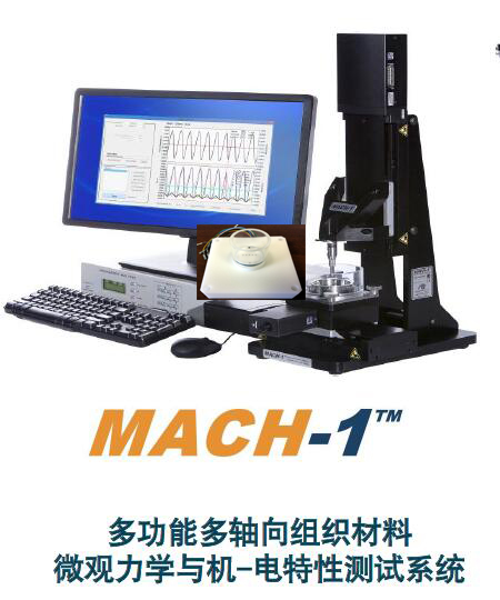
MachOne生物活体组织电位测试分析系统,材料机电特性测试分析系统,软骨等流动电位测试分析评估系统
型号:MachOne,
联系人:李先生
联系电话:18618101725
品牌:BMM
世联博研公司提供2款加拿大BMM 品牌的机电特性测试分析系统:
MachOne可以在压缩期间同时测量电势分布,系统可以活体在体测试软骨、骨的压电流动电位。
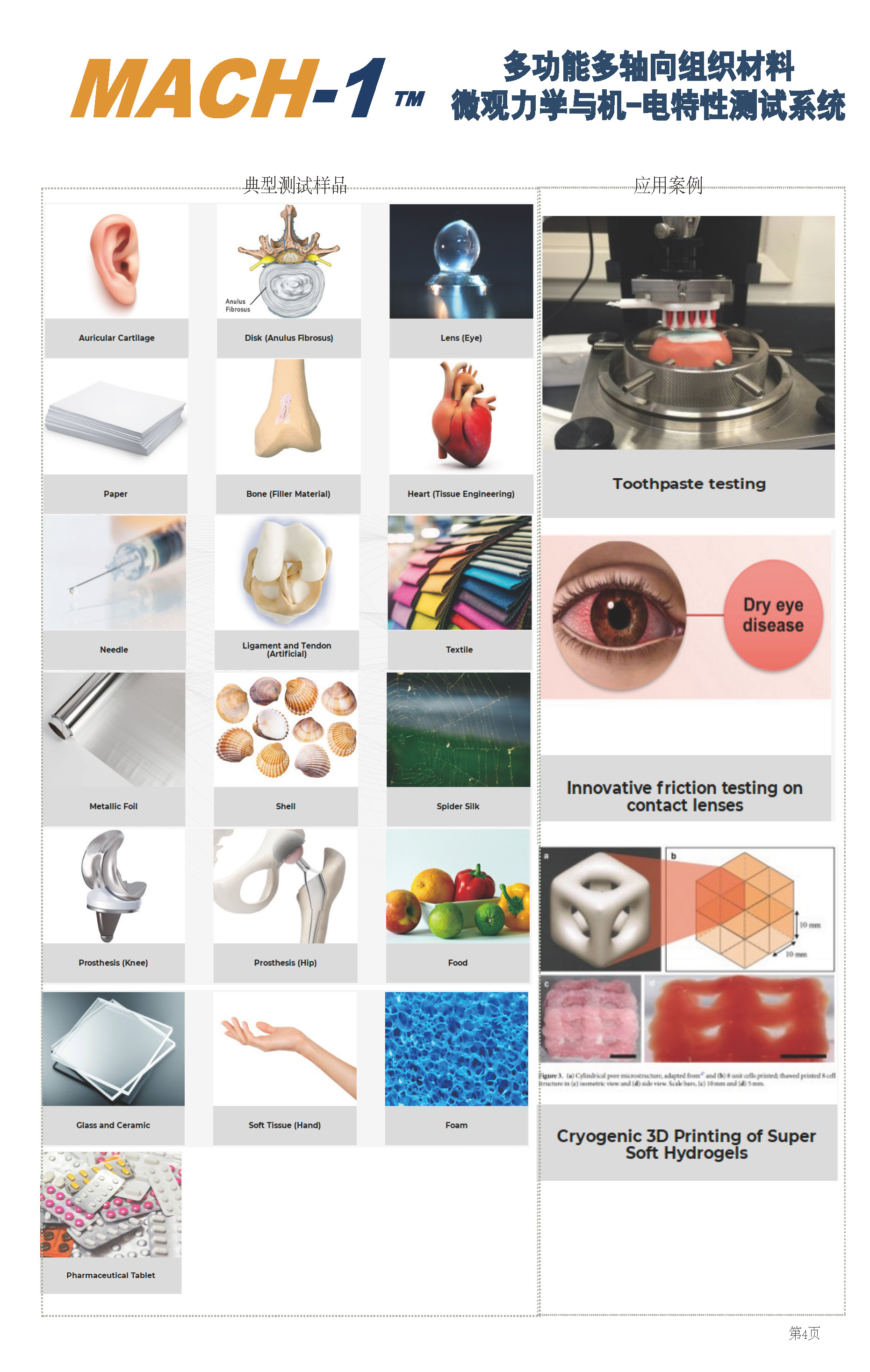
1、MachOne多功能组织材料生物力学特性、电位分布测试分析表征系统
-多载荷多物理场耦合微观力学性能原位测试系统
该BMM 材料/组织机电耦合效应特性测试仪(机械力偶联电位测试分析),可带电活性组织或材料进行机械力偶联电位测试分析,比如关节软骨、带电水凝胶等活组织压缩期间同步进行电位测试,可集成3D轮廓表面形貌表征、拉伸、压缩、三弯曲、四点弯曲、扭力、剪切、摩擦磨损、电特性等各种力电多物理场测试。 能对极软、极硬组织材料进行精密可靠的机械刺激和表征。允许表征的机械性能包括刚度、强度、模量、粘弹性、塑性、硬度、附着力、肿胀和松弛位移控制运动各种机械特性
特点
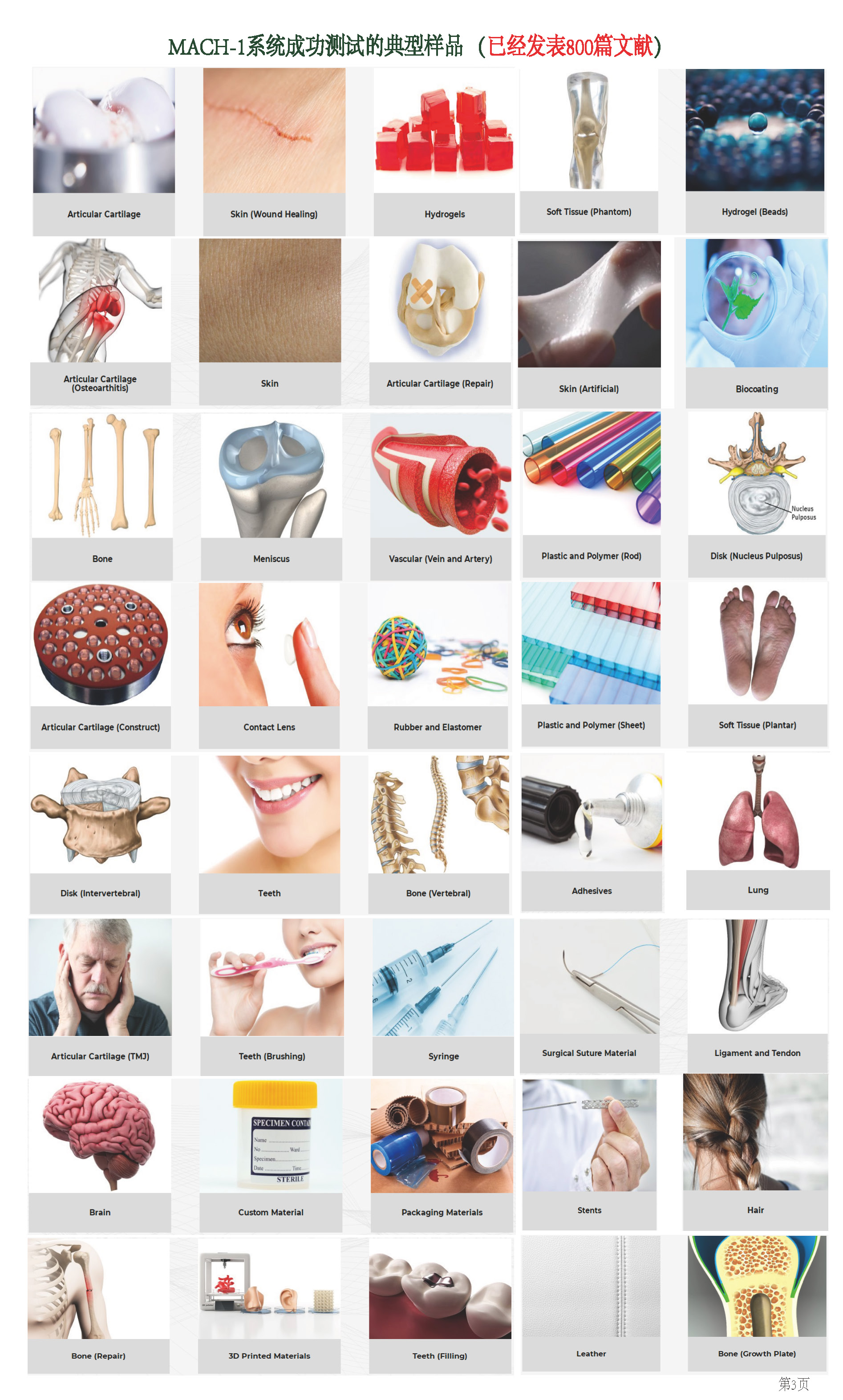
1、适用样品范围广:
1.1、从骨等硬组织材料到脑组织、眼角膜等软组织材料
1.2、从粗椎间盘的样品到j细纤维丝
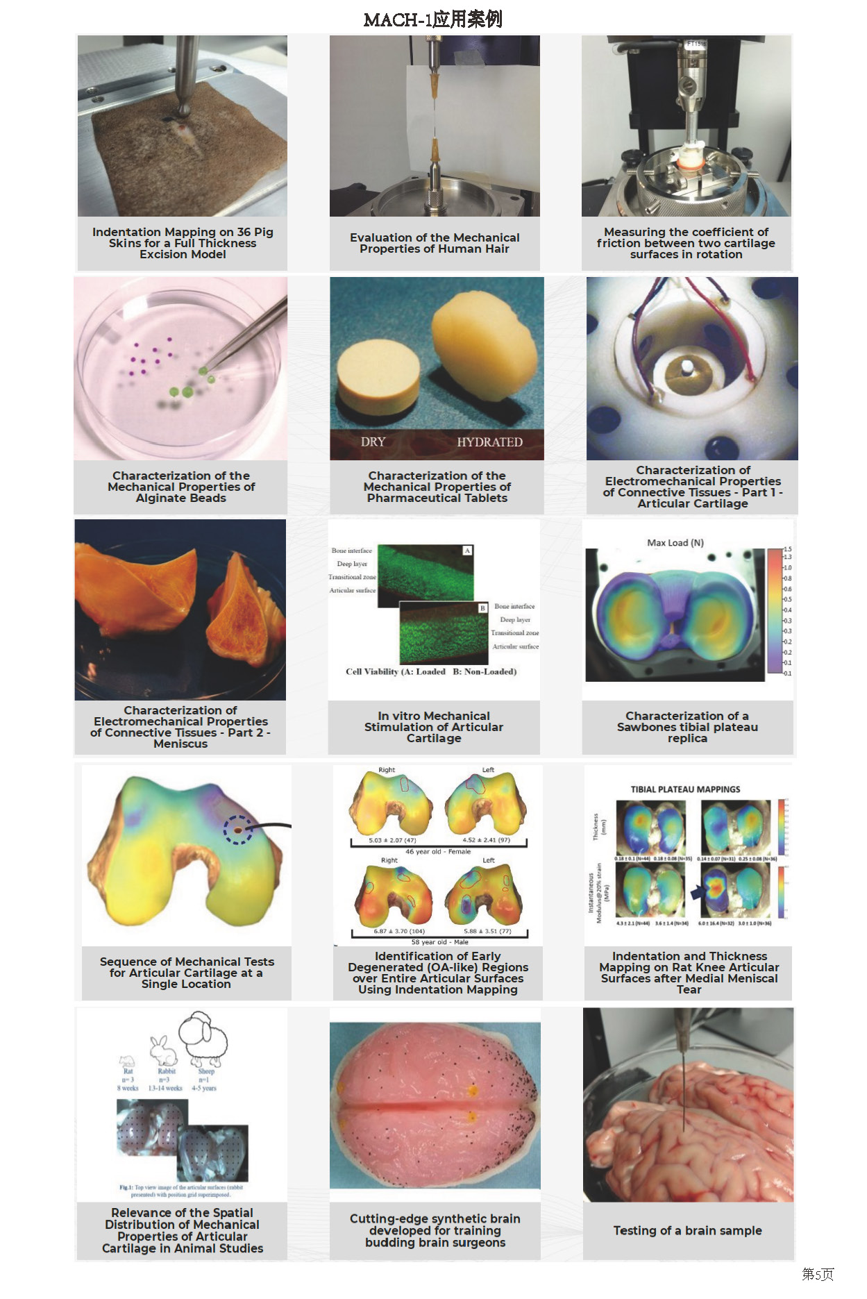
2、通高量压痕测试分析
2.1、三维法向压痕映射非平面样品整个表面的力学特性
2.2、48孔板中压痕测试分析
3、力学类型测试分析功能齐
模块化集成压缩、张力、剪切、摩擦、扭转、穿刺、摩擦和2D/3D压痕、3D表面轮廓、3D厚度等各种力学类型支持,微观结构表征及动态力学分析研究
4、高分辨率:
4.1、位移分辨率达0.1um
4.2、力分辨率 达0.025mN
5、 行程范围广:50-250mm
6、体积小巧、可放入培养箱内
7 、高变分辨率成像跟踪分析
8、多轴向、多力偶联刺激
9、活性组织电位分布测试分析
10、产品成熟,文献量达 上千篇
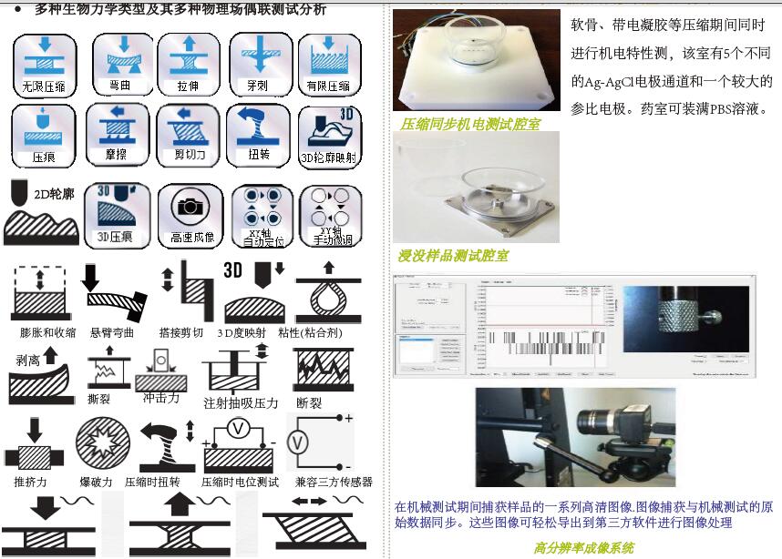
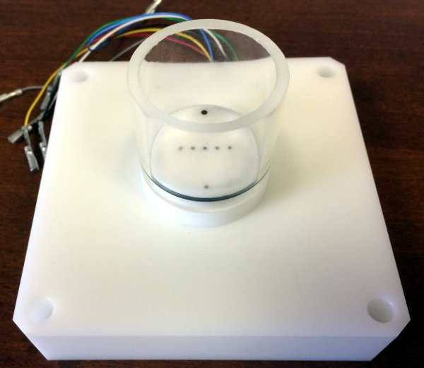
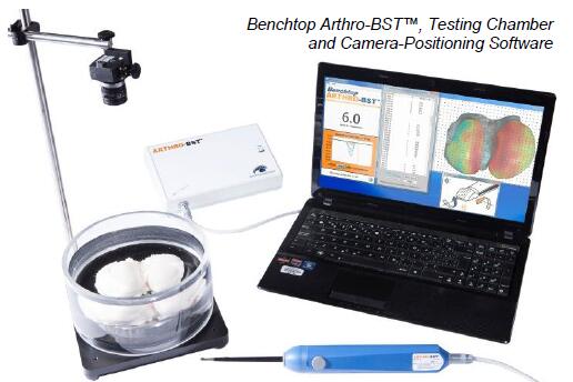
机电测试室如下:
该腔室设计用于在机电活性材料(例如结缔组织,带电水凝胶等)压缩期间同步测量电势分布。 该腔室具有5个差分Ag-AgCl电极通道和一个较大的参比电极。 圆形电极直径为1毫米,它们之间(中心到中心)的距离为4毫米。 可根据要求提供定制的几何形状。 腔室可以充满PBS溶液。
点与特点
该附件的设计便于清洁和消毒。
此零件设计坚固,可保持其完整性多年
设计用于与生理盐水溶液接触
易于组装和安装在测试仪上
由透明材料制成,方便组装
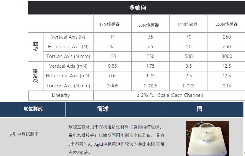
技术指标:
材质:Ag-AgCl(电极)直径:1 mm(电极)
间距:4 mm(中心到中心)(电极)
文献案例:
1、结缔组织的机电性能表征-
CHARACTERIZATION OF ELECTROMECHANICAL PROPERTIES OF CONNECTIVE TISSUES - PART 1 - ARTICULAR CARTILAGE
In this case study, the MachOne mechanical tester was used to determine the electromechanical properties of two connective tissues: the cartilage and the meniscus. Since tissue stiffness and compression-generated electric fields, or streaming potentials, are sensitive to the biochemical content of the tissue, the tester was coupled with an array of electrodes that can precisely measure these electric signals. The objective of this study was to establish links between biochemical composition and electromechanical properties (stiffness and streaming potentials) of normal and degenerated connective tissues. The first part of the study focuses on articular cartilage.
Since the articular cartilage extracellular matrix is primarily composed of negatively charged proteoglycans entrapped in a collagen network, there is an excess of mobile positive charges in the fluid. Compression of the cartilage produces streaming potentials via the separation of positively charged mobile ions relative to the fixed negatively charged proteoglycans.
Matched pairs of bovine cartilage/bone disks (3 mm in diameter) were cultured with and without a degradation agent (interleukin-1 or IL-1) for 11 days.
Electromechanical properties of the cartilage were evaluated by incorporating an array of electrodes into a testing chamber mounted on the MachOne. The MachOne is designed for various testing protocols including stress relaxation, sinusoidal displacement and creep in different configurations (confined and unconfined compressions, tension, indentation and bending). In this study, streaming potentials across the tissue surface are measured in unconfined compression geometry with a linear array of 8 platinum electrodes of 50 um diameter separated by 300 um. Electrodes 1 to 6 covered the 1.5 mm cartilage disk radius, and electrodes 7 and 8 were external to the cartilage in the bath. The testing chamber was filled
with physiological saline solution. A static compression offset of 100 μm was applied in a sequence of small step compressions of 20 μm at 2 μm/s. Dynamic sinusoidal tests were performed at frequencies of 1, 0.1 and 0.01 Hz with displacement amplitudes of 8, 4 and 2 μm. The streaming potential radial profile was constructed by the addition of each channel in the complex domain. Proteoglycan or glycosaminoglycan (GAG) content in the disk and content lost to the media during the culture was measured with the dimethylmethylene blue dye and spectrophotometry.
Figure 3 shows the GAG content in cartilage disks during the culture. The IL-1 treated disks lost 10, 20, 65 and 75% of their GAG content after 1, 4, 7 and 11 days of culture respectively. The MachOne is easy to use and its modular software permits a direct and rapid analysis of the dynamic sinusoidal test results. Figure 4 shows that dynamic stiffness was 3 times lower for the IL-1 treated disks compared to control after 11 days of culture.
The analysis of the stress relaxation test results showed that, at the end of the culture, the static stiffness was 4 times lower for degraded disks compared to control (0.3 MPa). Figure 5 shows the streaming potential profiles after 11 days of culture for normal and degraded cartilage. We observed that the profile potential gradient at the periphery for the IL-1 treated explants was reduced by half compared to control disks only one day after the beginning of the culture. The potential profile amplitude at the center of the disk was significantly lower for degraded disks compared to control explants after 7 days of culture.
The MachOne mechanical tester coupled with an array of electrodes can precisely determine the electromechanical properties of a large range of normal and degraded connective tissues.
2、结缔组织的机电性能表征-2部分-半月板
CHARACTERIZATION OF ELECTROMECHANICAL PROPERTIES OF CONNECTIVE TISSUES - PART 2 - MENISCUS
In this case study, the MachOne mechanical tester was used to determine the electromechanical properties of two connective tissues; the cartilage and the meniscus. Since tissue stiffness and compression-generated electric fields, or streaming potentials, are sensitive to the biochemical content of the tissue, the tester was coupled with an array of electrodes that can precisely measure these electric signals. The objective of this study was to establish links between biochemical composition and electromechanical properties (stiffness and streaming potentials) of normal and degenerated connective tissues. The second part of the study focuses on meniscus.
The meniscus is a tissue similar to cartilage but softer and less charged. In this case, the samples have been sectioned to five 3-mm disks (about 1-2 mm thick) from various specified regions. Dynamic stiffness under unconfined compression and the streaming potential distribution are measured. It should be noted that the streaming potential are around 10 times lower for meniscus than cartilage (refer to Part 1 of this case study). These results also indicate that the dynamic stiffness and the streaming potential profiles are higher on the cartilaginous surface of the inner part of the meniscus (disk 1 and 2) compared to middle zones (disks 3 and 5). It is expected that these results are directly related to the proteoglycan content of menisci.
The MachOne mechanical tester coupled with an array of electrodes can precisely determine the electromechanical properties of a large range of normal and degraded connective tissues.
2、软骨等流动电位测试分析评估系统
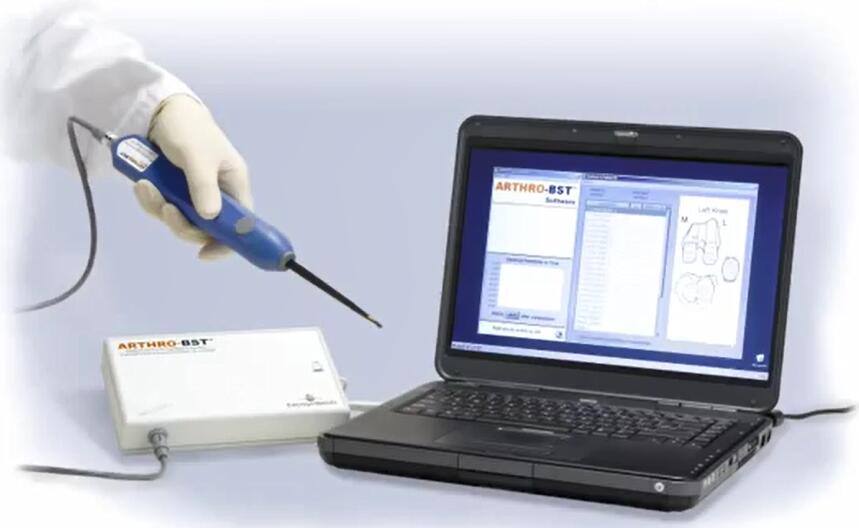
?是一种手持式医疗设备,设计用于关节镜手术,以评估关节软骨的功能特性。 通过压缩软骨表面,?测量流动电位并计算反映软骨健康状态的定量参数。
Benchtop ?可对关节软骨进行j确的wu损评估。 它计算了一个定量参数,反映了每个测量部位的关节软骨的生化成分和承重特性。 它是为有兴趣仅在离体关节软骨上使用该工具的研究人员开发的。 提供非wu菌一次性吸头,可配备配备高分辨率相机和相机定位软件的测试室,用于绘制大型骨软骨样本。
软骨研究
Benchtop ?在软骨修复领域提供了大量研究机会。 它非常宝贵:- 了解软骨疾病,包括软骨退化的潜在原因
- 开发新的治疗产品和软骨修复技术
- 可靠的骨关节炎动物模型的概念
- 创建各种物种的参考映射
工作原理:
台式?测量关节软骨的压缩诱导的流动电位。在温和压缩软骨表面期间使用压头测量这些电信号(参见下图)。te的压头设计包括覆盖有37个微电极阵列的球形表面。
该装置计算软骨机电活动的定量参数(QP),其对应于当其流动电位的总和达到100mV时与软骨接触的微电极的数量。高QP表示弱机电特性,反之亦然。 QP是可再现的,并且与施加的力和压头取向wu关。
健康的软骨正常流动电位:
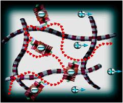
在正常软骨被压缩时,相对于包埋在胶原网络内的带负电荷的蛋白聚糖,间质液的流动取代带正电的移动离子。这产生称为流动电位的电位。
关节炎软骨低流动电位:
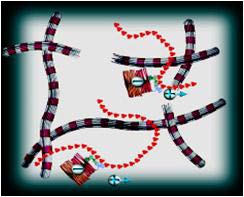
退化的软骨的特征在于蛋白多糖的丧失和胶原网络的减弱。由于相对于带负电的蛋白多糖,正离子的位移较小,因此关节炎软骨的压缩产生异常低的流动电位。
相关文献案例:
Literature ()