产品名称3D组织器官芯片模型, SynRAM 3D Inflammation Model ,SynRAM 3D炎症模型–一次分析即可可视化滚动,粘附和迁移
品牌synvivo
产品货号3D组织器官芯片模型, SynRAM 3D Inflammation Model ,SynRAM 3D炎症模型–一次分析即可可视化滚动,粘附和迁移
产品价格现货询价
联系人李先生
联系电话18618101725
产品说明

SynRAM 3D炎症模型

一次分析即可可视化滚动,粘附和迁移

SynVivo的SynRAM?3D炎症模型是为研究动态环境中的整个炎症途径而开发的。 SynVivo平台可提供生理上逼真的模型(包括流动和剪切),并能够实时跟踪轧制,粘附和迁移过程。该模型重建具有内皮细胞管腔的共培养组织和/或肿瘤细胞的组织切片。它已经成功地针对体内研究进行了验证,该研究显示出与滚动速度,粘附模式和迁移过程具有ji好的相关性(Lamberti等,2014; Soroush等,2016)。

SynRAM 3D炎症模型提供了一个现实的测试环境,其中包括:
微血管环境中的生理切应力
具有wanquan封闭腔的体内类血管形态
细胞间相互作用的共培养能力
单个实验的实时定量滚动,粘附和迁移数据
synram迁移测定原理图
芯片上白细胞内皮相互作用
SynRAM能够在一个实验中实时评估细胞相互作用,包括滚动,粘附和迁移通过多个细胞层,并代表与体内结果密切相关的数据。

SynRAM的创新设计克服了流动室或基于Transwell室的测定法固有的当前局限性。当前的流动室设计过于简单,缺乏微环境的规模和几何形状,并且wu法模拟迁移。同样,Transwell腔室也不能解释体内观察到的流体剪切力和大小/拓扑。另外,迁移的终点测量在Transwell腔室中wu法再现,并且不能提供实时可视化。

SynVivo的专有芯片设计范围从复杂的体内衍生的微血管网络(从数字化图像获得)到简化的里想化网络。复杂的体内网络会产生逼真的细胞组成和血管形态,从而导致剪切和流动条件发生变化。简化的里想化网络旨在重现细胞组成,恒定剪切力和流动条件。

在SynRAM器件中功能化的模型的示例
Synvivo Synram
实时可视化轧制,粘附,粘附和迁移
实时跟踪单细胞迁移
实时跟踪单细胞迁移
白细胞内皮相互作用
白细胞内皮相互作用
quan流明
血管内皮细胞芯片上的血管


产品购买选项

芯片:根据您的特定研究应用,您可以从IMN2径向SMN2微血管网络芯片配置中进行选择。

试剂盒:运行SynRAM分析所需的基本组件都可以以试剂盒形式购买。提供两种套件格式。

入门套件:shou次购买时请选择

10个SynRAM芯片(选择IMN2放射状或SMN2微血管网络芯片)
配件,包括油管,夹具,针头和注射器
气动灌注装置(灌注管路以除去空气时需要)
检测试剂盒:如果您以前购买过气动灌注设备,请选择此试剂盒格式

10个SynRAM芯片(选择IMN2径向或SMN2微血管网络芯片)
配件,包括油管,夹具,针头和注射器
用于开发SynRAM炎症模型的设备的示意图。顶端室(外部通道)用于培养血管(内皮细胞),而基底外侧室(中心腔)用于组织细胞培养。多孔结构使血管细胞与组织细胞之间可以进行通讯。


Bioinspired Microfluidic Assay for In Vitro Modeling of Leukocyte–Endothelium Interactions
Authors: G. Lamberti, B. Prabhakarpandian, C. Garson, A. Smith, K. Pant, B. Wang, and M.F. Kiani. Anal.
Chem., 2014, 86 (16), pp 8344–8351 DOI:10.1021/ac5018716

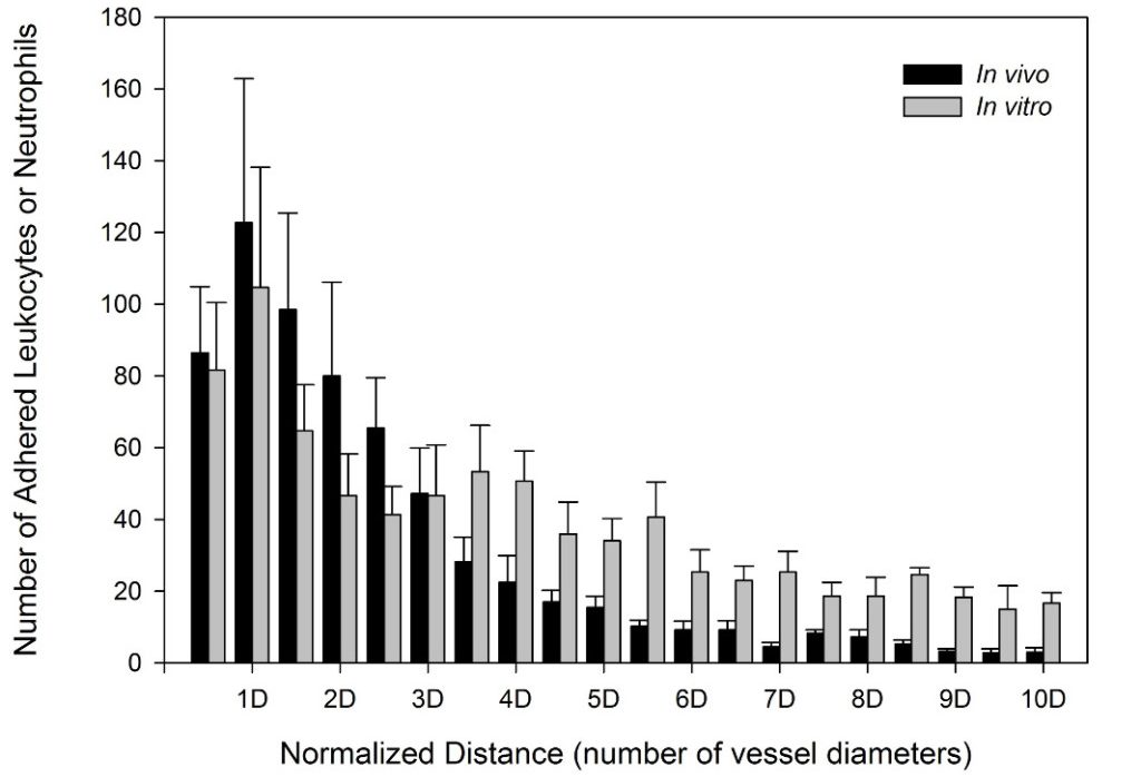
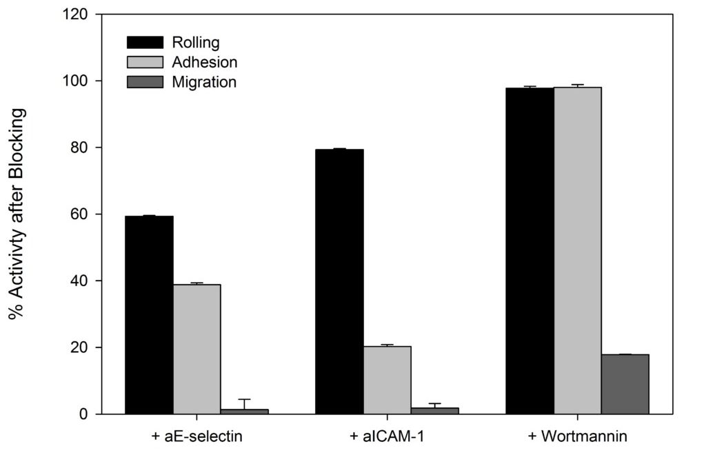
SynRAM microfluidic chips comprising of realistic microvascular networks were used to understand the role of classical inhibitors of individual steps of the leukocyte adhesion cascade. Experimental results matched very well with in vivo data highlighting the unique ability of the platform for real-time analysis of these dynamic events in a morphologically realistic environment (Lamberti et al 2014).

neutrophils charts
Rolling, adhesion, and migration of neutrophils in bMFA; migration of neutrophils (labeled with fluorescent dye) into the tissue compartment of bMFA after 120 min of continuous flow. (1 and 2) Solid arrows in the top right panels show a rolling neutrophil which (3) becomes adherent; dotted arrows in the top right panels show firmly adherent neutrophils. A neutrophil migrating from a vascular channel through the barrier into the tissue compartment over time (bottom right).
rolling velocity graph
Neutrophil rolling using SynRAM microfluidic chips is similar to leukocyte rolling in vivo; Box and whisker plots summarizes the comparison of leukocytes rolling velocity measured in vivo and in SynRAM chips and shows no significant difference (p=0.758; Mann-Whitney Rank Sum Test). The “+” marked in the box indicates the mean.
adhesion distance chart
Neutrophil adhesion in SynRAM microfluidic chips is similar to leukocyte adhesion in vivo; Distribution of the number of adhered leukocytes and neutrophils as a function of distance from the nearest bifurcation in vivo in mouse cremaster muscle model and in vitro in microfluidic chips, respectively. Both histograms are skewed to the left indicating that leukocytes and neutrophils preferentially adhere near bifurcations with the peak occurring at one vessel or channel diameter from the nearest bifurcation.

Investigation of the Effect of Blocking of Specific Steps of the Inflammation Pathway using Monoclonal Antibodies

Antibody blocking of specific steps in the adhesion/migration cascade downregulates other steps of the cascade; Monoclonal antibodies against E-selectin (aE-selectin), ICAM-1(aICAM-1), and PI3K (wortmannin) significantly reduced the number of rolling, adhering, and migrating neutrophils in SynRAM microfluidic devices.

activity after blocking chart
Antibody blocking of specific steps in the adhesion/migration cascade downregulates other steps of the cascade; monoclonal antibodies against E-selectin (aE-selectin), ICAM-1(aICAM-1), and PI3K (wortmannin) significantly reduced the number of rolling, adhering, and migrating neutrophils in bMFA. The numbers represent the percentage of activity after treating cells with the respective blockers in comparison to their corresponding control values (mean ± SEM; N = 3).

Elucidation of the Mechanism of Protein Kinase C delta (PKCδ) in Sepsis Related Inflammation Response

A Novel Microfluidic Assay Reveals a Key Role for Protein Kinase C δ in Regulating Human Neutrophil-Endothelium Interaction
Authors: Soroush F, Zhang T, King DJ, Tang Y, Deosarkar S, Prabhakarpandian B, Kilpatrick LE, Kiani MF.
J Leukoc Biol November 2016 100:1027–1035.

The SynRAM model was used to identify the underlying mechanism of Protein Kinase C delta (PKCδ) dependent neutrophil-endothelium interactions. These interactions have been found to play a significant role in the inflammatory response. They found that PKC? was a critical regulator of human neutrophil adhesion and migration through human endothelial cells during inflammation. This was validated by testing physiological fluid flow conditions of the entire inflammation process comprised of rolling, adhesion, and migration in real-time.

migration neutrophils chart
PKCδ inhibitor significantly reduces migration of neutrophils from the vascular channels, across the inflammed endothelium (treated with TNF-α for 4 or 24 hour), into the tissue compartment in response to fMLP mediated signaling compared to untreated controls.
sepsis detection chart
Immunohistochemical detection of myeloperoxidase (MPO) in representative lung tissue sections from 24 h post surgery. Few MPO-positive cells in Sham surgery. Sepsis induces the infiltration of numerous MPO-positive cells throughout the lung parenchyma. PKCδ-TAT Inhibitor significantly reduces sepsis-induced, MPO-positive cell numbers in the lung indicating decreased neutrophil migration.